Nursing Diagnosis and Nursing Intervention

Showing posts with label Appendicitis. Show all posts

Pathophysiology and Clinical Manifestation of Appendicitis

Pathophysiology and Clinical Manifestation of Appendicitis

Pathophysiology of Appendicitis

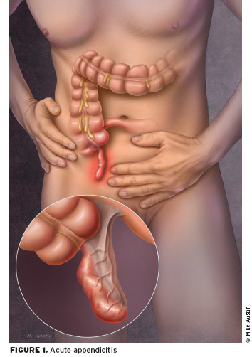
Appendix inflamed and had edema as a result of congestion, possibly by fecalith (hard mass of feces), tumor or a foreign object. Inflammatory process, increased intraluminal pressure that will impede lymph flow resulting in edema, diapedesis bacteria and ulceration of the mucosa cause upper abdominal pain or severe diffuse progressively, within a few hours, localized to the right lower quadrant of the abdomen. Finally, the inflamed appendix contains pus.

When mucus secretion continues, the pressure will continue to rise causing widespread inflammation and the resulting local peritoneum, causing pain under the right side is called acute suppurative appendicitis. If then the flow will be disrupted arterial wall infarction followed by a gangrenous appendix called gangrenous appendicitis. If the walls are already fragile perforated appendicitis rupture will occur. If all of the above process is slow, omentum and adjacent bowel will move toward an appendix to arise a local mass dsebut appendicular infiltrates. Inflammation of the appendix may be an abscess or disappear.

In children, shorter omentum and appendix are longer, thinner wall of the appendix. The situation is coupled with immune system becomes less ease of perforation. In older people perforation easily happen because there is an interruption of blood vessels (Mansjoer, 2000).

Clinical Manifestations of Appendicitis
  • Lower quadrant pain
  • Mild fever
  • Nausea and vomiting
  • Loss of appetite
  • Local tenderness at the point mc Burney
  • Tenderness off (or intesifikasi result of pain when pressure is released)
  • Signs rovsing can arise by doing palpoasi lower left quadrant which paradoksimal cause pain felt in the lower right quadrant
  • Abdominal distension due to paralytic ileus
  • The patient's condition deteriorates

Physical Examination, Laboratory, and Radiology for Acute Appendicitis

Physical Examination, Laboratory, and Radiology for Acute Appendicitis

There are several tests that can be performed by a medical team to determine and diagnose the presence of Appendicitis by patients. Among them is the physical examination, laboratory tests and radiology examinations:

1. Physical Examination of Acute Appendicitis

Physical Examination of Acute Appendicitis

In acute appendicitis, the observation would seem the swelling of the abdominal cavity which seemed to tighten the abdominal wall (distended). On palpation of the right lower abdominal area, often when pressed will feel pain when pressure is released and also will feel pain (Blumberg sign), which is the key to the diagnosis of acute appendicitis.

By the action of the right leg bent and thighs strong / leg high in the lift, the abdominal pain is getting worse. Suspicion of an inflammation of the appendix is ​​enhanced when an anal or vaginal and cause pain as well. Rectal temperature (rectal) temperatures are higher than the armpits, over longer support the existence of appendicitis.

2. Laboratory Examination of Acute Appendicitis

In laboratory tests of blood, which can be found is an increase of white blood cells (leukocytes) to about 10,000 - 18.000/mm3. If an increase is more than that, then it is likely the appendix had been perforated (ruptured).

3. Radiological Examination of Acute Appendicitis

Radiological Examination of Acute Appendicitis

Plain abdomen may show a fecalith. However, this examination is rarely helpful in diagnosing appendicitis. Ultrasonography (USG) is quite helpful in the diagnosis of appendicitis enforcement (71-97%), especially for pregnant women and children. The highest level of accuracy is by CT scan (93-98%). With a CT scan can be seen clearly picture the appendix.

Risk for Infection - Nursing Care Plan for Appendicitis

Risk for Infection - Nursing Care Plan for Appendicitis
Risk for Infection Appendicitis

Nursing Diagnosis Interventions for Appendicitis: Risk for Infection

Risk factors include:
  • Inadequate primary defense, perforation / rupture of the appendix; peritonitis; abscess formation.
  • Invasive procedures, surgical incisions.
Intervention:

Independent:
  • Monitor vital signs noticed fever, chills, sweating, mental changes, increased abdominal pain.
  • Do a good hand washing and aseptic wound care. Provide complete care.
  • See incision and bandage. Write down the characteristics and wound drainage / drain (if included), the erythema.
  • Provide appropriate information, be honest with the patient / parent close.
Collaboration
  • Take for example the drainage when indicated.
  • Give antibiotics, are as indicated.

Clinical Manifestations of Appendicitis

Clinical Manifestations of Appendicitis

  • Right lower quadrant pain felt and is usually accompanied by mild fever, nausea, vomiting, and loss of appetite.
  • Local tenderness at the point Mc.burney, when done pressure.
  • Tenderness may be encountered off.
  • The degree of muscle spasm and tenderness if there is constipation or diarrhea are not depending on the severity of the infection and the location of the appendix.
  • If the appendix behind the caecum circular, pain and tenderness can feel the lumbar region: when the tip is in the pelvis, these signs can be detected only by examination of the rectal examination.
  • Pain on defecation show ends appendix is near the rectum; pain during urination indicates that the tip of the appendix is close to the bladder or ureter.
  • There is stiffness in the bottom of the right testis muscles can occur.
  • Signs Rovsing can arise with left lower quadrant palpation paradoxical that causes pain felt in the lower right quadrant. If paralytic ileus, and the patient's condition deteriorated.
  • In elderly patients, the signs and symptoms of appendicitis can vary greatly. The signs can be very dubious, suggesting destruction of the bowel or other disease processes. Patients may have no symptoms until he suffered a ruptured appendix. The incidence of perforation of the appendix is higher in the elderly, because many of these patients do not seek medical care as soon as patients are younger.
Copyright © Nursing Diagnosis Intervention. All rights reserved. Template by CB | Published By Kaizen Template | GWFL | KThemes