Nursing Diagnosis and Nursing Intervention

Pathophysiology and Clinical Manifestation of Appendicitis

Pathophysiology and Clinical Manifestation of Appendicitis

Pathophysiology of Appendicitis

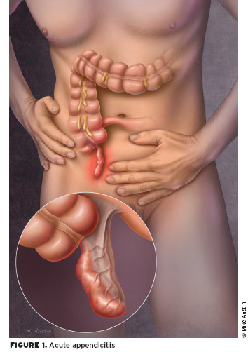
Appendix inflamed and had edema as a result of congestion, possibly by fecalith (hard mass of feces), tumor or a foreign object. Inflammatory process, increased intraluminal pressure that will impede lymph flow resulting in edema, diapedesis bacteria and ulceration of the mucosa cause upper abdominal pain or severe diffuse progressively, within a few hours, localized to the right lower quadrant of the abdomen. Finally, the inflamed appendix contains pus.

When mucus secretion continues, the pressure will continue to rise causing widespread inflammation and the resulting local peritoneum, causing pain under the right side is called acute suppurative appendicitis. If then the flow will be disrupted arterial wall infarction followed by a gangrenous appendix called gangrenous appendicitis. If the walls are already fragile perforated appendicitis rupture will occur. If all of the above process is slow, omentum and adjacent bowel will move toward an appendix to arise a local mass dsebut appendicular infiltrates. Inflammation of the appendix may be an abscess or disappear.

In children, shorter omentum and appendix are longer, thinner wall of the appendix. The situation is coupled with immune system becomes less ease of perforation. In older people perforation easily happen because there is an interruption of blood vessels (Mansjoer, 2000).

Clinical Manifestations of Appendicitis
  • Lower quadrant pain
  • Mild fever
  • Nausea and vomiting
  • Loss of appetite
  • Local tenderness at the point mc Burney
  • Tenderness off (or intesifikasi result of pain when pressure is released)
  • Signs rovsing can arise by doing palpoasi lower left quadrant which paradoksimal cause pain felt in the lower right quadrant
  • Abdominal distension due to paralytic ileus
  • The patient's condition deteriorates

Nursing Diagnosis Decreased Cardiac Output for Hyperthyroidism

Decreased Cardiac Output related to uncontrolled hyperthyroidism, hypermetabolism, increased cardiac workload.

Patients objective / evaluation criteria;
  • Maintain adequate cardiac output according to the needs of the body

characterized by:
  • Stable vital signs,
  • normal peripheral pulses,
  • normal capillary refill,
  • good mental status,
  • no dysrhythmias

Nursing Intervention:

Independent
  • Monitor vital signs. Note the magnitude of the pressure pulse.
  • Check / meticulous possibility complained of chest pain patients.
  • Assess pulse / heart rate while the patient sleeps.
  • Auscultation of heart sounds, note the extra heart sounds, a gallop rhythm and a systolic murmur.
  • ECG monitor, record or note rate or in cardiac rhythm and the presence of dysrhythmias
  • Observation of signs and symptoms of severe thirst, dry mucous membranes, weak pulse, slow capillary refill, decreased urine output, and hypotension
  • Note adnya history of asthma / bronkokontriksi, pregnancy, sinus bradycardia / heart block progress to heart failure
Collaboration
  • Give fluids through IV as indicated
  • Give medications as indicated:
  • Monitor the results of lab tests: serum potassium, serum calcium, sputum culture
  • Perform regular ECG monitoring
  • Give oxygen as indicated
  • Prepare for surgery

Meningitis Definition, Etiology and Clinical Manifestations

Definition of Meningitis

Meningitis is inflammation of the meninges (the membranes that surround the brain and spinal cord) and is caused by a virus, bacteria or fungus organs (Smeltzer, 2001).

Meningitis is an infection of the fluid of the brain with inflammatory piamater, arachnoid and in a milder degree of the brain and spinal cord tissues were superficial. (Neurology capita selekta, 1996)

Meningitis is an inflammation of the arachnoid and pia mater (lepto meningens) of the brain and spinal cord. Bacteria and viruses are the most common cause of meningitis, although fungi can also cause. Bacterial meningitis is more common. Early detection and treatment will give more better results according to Revelation Widagdo et al (2008:105)

Etiology of Meningitis

Meningitis caused by a virus is generally harmless, will recover without specific treatment and care. But bacterial meningitis can lead to serious conditions, such as brain damage, hearing loss, lack of ability to learn, can even cause death. While meningitis is caused by a fungus is very rare, this type generally affects people with damaged immune (immune system) as in patients with AIDS.

Bacteria that can cause meningitis attack include:
1. Streptococcus pneumoniae (pneumococcus).
These bacteria are the most common cause of meningitis in infants or children. This type of bacteria can also cause pneumonia, ear and nasal cavity (sinus).

2. Neisseria meningitidis (meningococcus).
This bacterium is the second most after Streptococcus pneumoniae meningitis caused by an infection of the upper respiratory tract and then the bacteria enter the bloodstream.

3. Haemophilus influenzae (Haemophilus).
Haemophilus influenzae type b (Hib) is a type of bacteria that can also cause meningitis. This type of virus as the cause upper respiratory infections, middle ear and sinuses. Vaccine (Hib vaccine) has shown a decrease in the number of cases of meningitis caused by these bacteria.

4. Listeria monocytogenes (listeria).
This is one type of bacteria that can cause meningitis. These bacteria can be found in many places, in the dust and in contaminated food. Food is usually a type of cheese, hot dogs and bacon sandwich which is derived from the bacterium local animal (pet).

5. Other bacteria that can also cause meningitis are Staphylococcus aureus and Mycobacterium tuberculosis.

Clinical Manifestations of Meningitis
  • Early in the disease, fatigue, changes in power to remember, change in behavior
  • In accordance with the rapid course of the disease the patient becomes stuporous
  • Headache
  • Sore muscle pain
  • Pupillary reaction to light. Photofobia when light is directed at the patient's eye.
  • Dysfunction of the nerves III, IV, VI
  • Motor movement at the beginning of the disease is usually normal and common in the advanced stages of hemiparesis, hemiplagia, and decreased muscle tone
  • Reflex positive Brudzinski and Kernig reflex
  • Nausea
  • Vomiting
  • Tachycardia
  • Convulsions
  • Patients feel fear and anxiety

Risk for Injury - Nursing Care Plan Meningitis


Nursing Diagnosis for Meningitis: Risk for Injury related to general weakness.

The expected outcomes / evaluation criteria pediatric patients: No seizures or comorbidities or other injury.

intervention
a. Monitor the spasms / twitching of the hands, feet and mouth or other facial muscles.
Rational: reflecting on the CNS in general irritation that require immediate evaluation and possible intervention to prevent complications.

b. Provide security for patients by providing assistance on the bed and keep the barrier remained in place and attach the plastic artificial airway or soft rolls and a suction bulb.
Rationale: protect patients when seizures. Note; enter the airway assistance / soft rolls if only jaw relaxation, not forced to enter as his teeth shut and soft tissue will be damaged.

c. Maintain bed rest during the acute phase. Move. Moving with the help of corresponding improvement in the situation.
Rational: reducing the risk of falls / trauma case vertigo, syncope or ataxia.

d. Give medication as indicated as phenytoin (Dilantin), diazepam, phenobarbital.
Rational: an indication for the treatment and prevention of seizures. Records: Phenobarbital may cause respiratory and sedative defresi and mask the signs / symptoms of increased ICP.

Retropharyngeal Abscess Prevention and Treatment

Retropharyngeal abscess Prevention and Treatment

Definition of retropharyngeal abscess

Retrofaringeal abscess is a pus accumulation in the back of the throat tissues.

Cause of retropharyngeal abscess
  • Abscesses are usually caused by streptococcal infection from the tonsils, throat, sinuses, adenoids, nasal or middle ear.
  • Sometimes an injury to the back of the throat caused by thorn fish can also cause abscesses retrofaringeal.
  • Although rare, retrofaringel abscess can also be caused by tuberculosis.
  • Retrofaringeal abscess usually affects children aged less than 5 years.
  • Networking on the back of the throat allows the formation of children's cavity containing pus (which this does not occur in adults).
  • Infection in these areas can occur during or shortly after by a bacterial throat infection.

Symptoms of retropharyngeal abscess

Symptoms include:
  • History of sore throat, nasal infection or tooth abscess
  • High fever
  • Severe sore throat
  • Swollen neck lymph nodes
  • Difficulty swallowing
  • Salivating
  • Respiratory Disorders
  • Intercostal retraction (withdrawal of muscles between the ribs when people try so hard to breathe)
  • Stridor (harsh breathing sound).

Complications of retropharyngeal abscess
  • Bleeding around the abscess
  • Rupture of the abscess into the airways (which can cause airway obstruction)
  • Pneumonia
  • The spread of infection to the chest.
Diagnosis of retropharyngeal abscess
  • Diagnosis is based on symptoms and physical examination.
  • X-rays or a CT scan of the neck showed a cavity containing pus between the throat and cervical spine.
  • Blood tests showed an increase in the number of white blood cells.
  • Breeding throat mucus can indicate the presence of the causative organism.

Treatment of retropharyngeal abscess
  • To overcome infection drainage surgery (to remove the pus) and high doses of antibiotics given intravenously.

Prevention of retropharyngeal abscess
  • Diagnosis and treatment in pharyngitis and nasopharyngeal infection usually can prevent abscess retrofaringeal.

Physical Examination, Laboratory, and Radiology for Acute Appendicitis

Physical Examination, Laboratory, and Radiology for Acute Appendicitis

There are several tests that can be performed by a medical team to determine and diagnose the presence of Appendicitis by patients. Among them is the physical examination, laboratory tests and radiology examinations:

1. Physical Examination of Acute Appendicitis

Physical Examination of Acute Appendicitis

In acute appendicitis, the observation would seem the swelling of the abdominal cavity which seemed to tighten the abdominal wall (distended). On palpation of the right lower abdominal area, often when pressed will feel pain when pressure is released and also will feel pain (Blumberg sign), which is the key to the diagnosis of acute appendicitis.

By the action of the right leg bent and thighs strong / leg high in the lift, the abdominal pain is getting worse. Suspicion of an inflammation of the appendix is ​​enhanced when an anal or vaginal and cause pain as well. Rectal temperature (rectal) temperatures are higher than the armpits, over longer support the existence of appendicitis.

2. Laboratory Examination of Acute Appendicitis

In laboratory tests of blood, which can be found is an increase of white blood cells (leukocytes) to about 10,000 - 18.000/mm3. If an increase is more than that, then it is likely the appendix had been perforated (ruptured).

3. Radiological Examination of Acute Appendicitis

Radiological Examination of Acute Appendicitis

Plain abdomen may show a fecalith. However, this examination is rarely helpful in diagnosing appendicitis. Ultrasonography (USG) is quite helpful in the diagnosis of appendicitis enforcement (71-97%), especially for pregnant women and children. The highest level of accuracy is by CT scan (93-98%). With a CT scan can be seen clearly picture the appendix.

Risk for Infection - Nursing Care Plan for Appendicitis

Risk for Infection - Nursing Care Plan for Appendicitis
Risk for Infection Appendicitis

Nursing Diagnosis Interventions for Appendicitis: Risk for Infection

Risk factors include:
  • Inadequate primary defense, perforation / rupture of the appendix; peritonitis; abscess formation.
  • Invasive procedures, surgical incisions.
Intervention:

Independent:
  • Monitor vital signs noticed fever, chills, sweating, mental changes, increased abdominal pain.
  • Do a good hand washing and aseptic wound care. Provide complete care.
  • See incision and bandage. Write down the characteristics and wound drainage / drain (if included), the erythema.
  • Provide appropriate information, be honest with the patient / parent close.
Collaboration
  • Take for example the drainage when indicated.
  • Give antibiotics, are as indicated.

Clinical Manifestations of Appendicitis

Clinical Manifestations of Appendicitis

  • Right lower quadrant pain felt and is usually accompanied by mild fever, nausea, vomiting, and loss of appetite.
  • Local tenderness at the point Mc.burney, when done pressure.
  • Tenderness may be encountered off.
  • The degree of muscle spasm and tenderness if there is constipation or diarrhea are not depending on the severity of the infection and the location of the appendix.
  • If the appendix behind the caecum circular, pain and tenderness can feel the lumbar region: when the tip is in the pelvis, these signs can be detected only by examination of the rectal examination.
  • Pain on defecation show ends appendix is near the rectum; pain during urination indicates that the tip of the appendix is close to the bladder or ureter.
  • There is stiffness in the bottom of the right testis muscles can occur.
  • Signs Rovsing can arise with left lower quadrant palpation paradoxical that causes pain felt in the lower right quadrant. If paralytic ileus, and the patient's condition deteriorated.
  • In elderly patients, the signs and symptoms of appendicitis can vary greatly. The signs can be very dubious, suggesting destruction of the bowel or other disease processes. Patients may have no symptoms until he suffered a ruptured appendix. The incidence of perforation of the appendix is higher in the elderly, because many of these patients do not seek medical care as soon as patients are younger.

Clinical Manifestations of Acute Gastritis and Chronic Gastritis

Clinical Manifestations of Acute Gastritis and Chronic Gastritis
Acute Gastritis and Chronic Gastritis

Clinical Manifestations of Acute Gastritis and Chronic Gastritis

Acute Gastritis

Acute Gastritis is very varied, ranging from very mild, asymptomatic, up to very heavy, which can lead to death. In the case of very heavy, very prominent symptoms are:
  1. Hematemetis and melena that can last a very great shock to occur due to blood loss.
  2. In most cases, symptoms are very mild and even asymptomatic. Complaints such as pain that arises in the gut, usually mild and can not be appointed to the appropriate location.
  3. Sometimes accompanied by nausea and vomiting.
  4. GI bleeding is often the only symptom.
  5. In the case of very light bleeding, manifests as occult blood in the stool and will be found in physical signs of deficiency anemia with no apparent etiology.
  6. On physical examination found no abnormalities except usually those with severe bleeding, causing signs and symptoms of hemodynamic disorders as real as hypotension, pallor, cold sweat, tachycardia until the disturbance of consciousness.
Chronic Gastritis
  1. Varied and unclear.
  2. Feeling of fullness, anorexia.
  3. Epigastric distress that is not real.
  4. Early satiety.

Pathophysiology of Chronic Gastritis

Pathophysiology of Chronic Gastritis
Chronic gastritis can be classified as type A or type B.

Type A (often referred to as autoimmune gastritis) resulting from changes in parietal cells, leading to atrophy and cell infiltration. It is associated with autoimmune diseases, such as pernicious anemia and occurs in the fundus or corpus of the stomach.

Type B (sometimes referred to as Helicobacter pylori, also known as H. pylori) is associated with the bacteria H. pylori, dietary factors such as heat or spicy drink, use drugs and alcohol, smoking or reflux of intestinal contents into the stomach. H. Pylori includes bacteria that are not acid resistant, but this kind of bacteria are able to secure himself in the mucosal lining of the stomach. The presence of this bacterium in gastric mucosa of the stomach lining causing weakening and brittle so that stomach acid can penetrate the layer. Thus both gastric acid and the bacteria causing wound or ulcer. The immune system will respond to bacterial infection H. Pylori by sending beads of leukocytes, killer T cells, and other infection-fighting. However, it is not able to fight infection H. The reason pylori can not penetrate the stomach lining. But also can not be removed so that the immune response continues to increase and grow. Polymorph die and release of superoxide radicals damaging compounds in the cells lining the stomach. Extra nutrients are sent to strengthen the leukocyte cells, but nutrition is also a source of nutrients for H. Pylori. Finally, the state of the damaged epithelium of the stomach, forming a superficial ulceration and can cause hemorrhage (bleeding). Within a few days gastritis and even peptic ulcers are formed.
Copyright © Nursing Diagnosis Intervention. All rights reserved. Template by CB | Published By Kaizen Template | GWFL | KThemes