Nursing Diagnosis and Nursing Intervention

Nursing Diagnosis related to Fluid and Electrolyte

Fluid and Electrolyte

1. Deficient Fluid volume: less than body requirements related to excessive fluid output.

Intervention:
  • Observation of vital signs.
  • Observed signs of dehydration.
  • Measure the input and output of fluid (fluid balance).
  • Provide and encourage families to give drink plenty of approximately 2000 - 2500 cc per day.
  • Collaboration with physicians in the delivery of fluid therapy, electrolyte laboratory examination.
  • Collaboration with a team of nutrition in low-sodium fluid administration.


2. Risk for Deficient fluid volume related to insufficient fluid intake, excessive discharge (vomiting / nausea).

Intervention:
  • Record the number of vomiting and bleeding characteristics.
  • Assess vital signs (BP, pulse, temperature).
  • Monitor fluid intake and output.
  • Elevate the head for taking medication.
  • Give saturated liquid / soft if the input starts again, avoid caffeinated and carbonated beverages.
  • Maintain bed rest.
  • Collaboration with fluid administration as indicated.

3. Risk for ineffective airway clearance related to the operative incision site.

Intervention:
  • Give analgesics as prescribed.
  • Fixation incision with both hands or a pillow to help patients when they cough.
  • Encourage the use of Incentive spirometer if there is an indication.
  • Help and encourage early ambulation.
  • Help the patient to change positions frequently.

4. Disturbed Body Image related to changes in appearance secondary to loss of body parts.

Intervention:
  • Encourage the patient to express feelings, especially about the thoughts, feelings, views of self. Rational: Helping patients to be aware of unusual feelings.
  • Note withdrawing behavior. Increased dependency, manipulation or not involved in treatment. Rational: Alleged problems in assessment can require follow-up evaluation and more rigorous therapy.
  • Maintain a positive approach during maintenance activities. Rational: Help the patient / person closest to accept changes in their own bodies and feel good about themselves.

Nursing Diagnosis for Urinary / Bowel Elimination : Diarrhea, Constipation

Nanda Nursing Diagnosis for Urinary / Bowel Elimination : Diarrhea, Constipation


1. Alteration in Bowel Elimination : Diarrhea

Intervention:
  • Help need for defecation (if bed rest to prepare the necessary tools near the bed, attach the curtains and immediately dispose of faeces after defecation).
  • Increase / maintain fluid intake by mouth.
  • Teach about the foods and drinks that can worsen / precipitate diarrhea.
  • Observation and record the frequency of defecation, fecal volume and characteristics.
  • Observation fever, tachycardia, lethargy, leukocytosis, decreased serum protein, anxiety and lethargy.
  • Collaboration of appropriate medication therapy program (antibiotics, anticholinergics, corticosteroids).


2. Alteration in Bowel Elimination : Constipation

Intervention:
  • Encourage lots of drinking with ambulation dinikolab laxative administration.
  • Rationalization:
  • Many drinks can help dissolve the stool with ambulation reduce constipation.
  • Formation of stools soft launch.

3. Alteration in Bowel Elimination: Constipation related to neurological disorders of the intestine and rectum.

Intervention:
  • Auscultation of bowel sounds, note the location and characteristics. Rational: bowel sounds may be absent during spinal shock.
  • Observe for abdominal distention.
  • Note the presence of complaints of nausea and want to vomit, pairs of NGT. Rational: gantrointentinal and gastric bleeding may occur due to trauma and stress.
  • Provide a balanced diet high in calories and protein; Liquid. Rational: improving stool consistency.
  • Give laxatives to order. Rational: stimulate the intestines.

4. Altered Urinary Elimination related to the drainage of urine.

Intervention:
  • Assess urine drainage system immediately.
  • Assess the adequacy of urine output and drainage system patency.
  • Use aseptic procedures and washing hands when providing care and action.
  • Maintain a closed urine drainage system.
  • If irrigation is needed and prescribed, do this action carefully using sterile saline.
  • Assist patients in the mobilization.
  • Observation of color, smell and consistency of urine volume.
  • Reduce trauma and manipulation of catheters, drainage system and urethra.
  • Clean the catheter carefully.
  • Maintain adequate fluid intake.

5. Impaired Urinary Elimination

Intervention:
  • Observation of the bladder.
  • Encourage regular bowel movements.
  • Give warm compresses.
  • Rationalization:
  • The content of urinary maintain contractions or uterine involution.
  • Urine retained causes infection.
  • Relaxation springter urine.

6. Altered Urinary Elimination related to paralysis of the urinary condition.

Intervention:
  • Assess the pattern of urination, and record urine output per hour.
  • Rationale: determine kidney function.
  • Palpation of the possibility of bladder distension.
  • Instruct the patient to drink a 2000 cc / day.
  • Rationale: helps maintain kidney function.
  • Attach the catheter Dower.
  • Rational assist the process of urine.

7. Constipation
Intervention:
  • Observation bowel sounds periodically.
  • Suggest to increase fluid intake at least 2 liters a day when no contra indications.
  • Increase activity on a regular basis.
  • For the provision of appropriate therapy, investigation is needed.
  • Dietis team collaboration for the provision of a balanced diet and high in fiber.

Causes of Diarrhea : Virus, Bacteria, Protozoa and Helminth

Causes of Diarrhea : Virus, Bacteria, Protozoa and Helminth
Cause of Diarrhea: (Tantivanich, 2002; Sirivichayakul, 2002; Pitisuttithum, 2002)

1. Virus:
Is the highest cause of acute diarrhea in children (70-80%). Some types of viruses that cause acute diarrhea:
  • Rotavirus serotypes 1,2,8, and 9: in humans. Serotype 3 and 4 were found in animals and humans. And serotypes 5,6, and 7 were found only in animals.
  • Norwalk virus: present in all ages, generally due to food borne or water borne transmission, and the transmission can also occur person to person.
  • Astrovirus, found in children and adults
  • Adenovirus (type 40, 41)
  • Small bowel structured viruses
  • Cytomegalovirus

2 Bacteria:
    Enterotoxigenic E. coli
  • Enterotoxigenic E. coli (ETEC). Have two important virulence factor is a factor that causes bacterial colonization is attached to the enterocytes of the small intestine and enterotoxin (heat labile (HL) and heat stabile (ST) which causes the secretion of fluid and electrolytes that produce watery diarrhea. ETEC causes no damage to brush border or invade the mucosa.
  • Enterophatogenic E. coli (EPEC). The mechanism of diarrhea is not clear. Found the process of attachment of EPEC to intestinal epithelial damage of membrane micro-villi which would disturb the surface absorption and disaccharidase activity.
  • Enteroaggregative E. coli (EAggEC). These bacteria are strongly attached to the mucosa of the small intestine and causes typical morphological changes. How does the mechanism of the onset of diarrhea is still unclear, but it may play a role cytotoxins.
  • Enteroinvasive E. coli (EIEC). In serologic and biochemical similar to Shigella. Such as Shigella, EIEC penetrate and multiply within colonic epithelial cells.
  • Enterohemorrhagic E. coli (EHEC). EHEC producing verocytotoxin (VT) 1 and 2, which is also called Shiga-like toxin that causes diffuse edema and bleeding in the colon. In children often progress to hemolytic-uremic syndrome.
  • Shigella spp. Shigella invade and multiply within colonic epithelial cells, causing cell death and the onset of mucosal ulceration. Shigella rarely enter into the bloodstream. Virulence factors including: smooth cell-wall lipopolysaccharide antigen and endotoxin activity has helped the process of invasion and toxin (Shiga toxin and Shiga-like toxin) that are cytotoxic and neurotoxic and may cause watery diarrhea.
  • Campylobacter jejuni (Helicobacter jejuni). Humans become infected through direct contact with animals (birds, dogs, cats, sheep and pigs) or with animal feces through contaminated food such as chicken and water. Sometimes the infection can be spread through direct person to person contact. C.jejuni may cause diarrhea by invasion into the small intestine and colon great.There 2 types of toxin produced, the heat-labile cytotoxin and enterotoxin. Histopathological changes that occur similar to the process of ulcerative colitis.
  • Vibrio cholerae 01 and V.cholerae 0139. water or food contaminated with this bacteria will transmit cholera. Through person to person transmission is rare.
  • V.cholerae attached and proliferated on the mucosa of the small intestine and produces an enterotoxin that causes diarrhea. Cholera toxin is very similar to the heat-labile toxin (LT) of ETEC. The last discovery of the existence of other enterotoxin that has its own characteristics, such as the accessory cholera enterotoxin (ACE) and zonular occludens toxin (ZOT). Both of these toxins cause fluid secretion into the intestinal lumen.
  • Salmonella (non-typhoid). Salmonella can invade intestinal epithelial cells. Produced enterotoxin causing diarrhea. If there is damage that causes mucosal ulcers, bloody diarrhea will occur.

3. Protozoa:
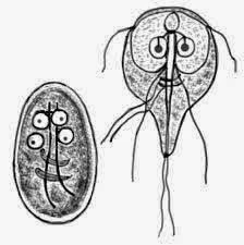
    Giardia lamblia
  • Giardia lamblia. This parasite infects the small intestine. Patogensis mechanism remains unclear, but is believed to affect the absorption and metabolism of bile acids. Transmission through the fecal-oral route. Host-parasite interactions is affected by age, nutritional status, endemicity, and immune status. Areas with high endemicity, giardiasis can be asymptomatic, chronic, persistent diarrhea with or without malabsorption. In areas with low endemicity, outbreaks can occur within 5-8 days after exposure to the manifestation of acute diarrhea is accompanied by nausea, epigastric pain and anorexia. Sometimes encountered malabsorption with faty stools, abdominal pain and bloated.
  • Entamoeba histolytica. Dysentery amoeba prevalence varies, but its spread throughout the world. The incidence increases with age, and teranak in adult males. Approximately 90% of asymptomatic infections caused by non-pathogenic E.histolytica (E.dispar). Symptomatic amebiasis can be mild and persistent diarrhea to fulminant dysentery.
  • Cryptosporidium. In developing countries, cryptosporidiosis 5-15% of cases of diarrhea in children. The infection is usually symptomatic and asymptomatic infants in older children and adults. Clinical symptoms of acute diarrhea with watery type of diarrhea, usually mild and self-limited. In people with impaired immune systems such as AIDS patients, cryptosporidiosis is a reemerging disease with more severe diarrhea and resistant to some antibiotics.
  • Microsporidium spp
  • Isospora belli
  • Cyclospora cayatanensis

4. Helminths:
  • Strongyloides stercoralis. Abnormalities in intestinal mucosa caused by adult worms and larvae, causing diarrhea.
  • Schistosoma spp. The blood worms cause abnormalities in various organs including the intestinal manifestations, including diarrhea and intestinal bleeding.
  • Capilaria philippinensis. This worm is found in the small intestine, especially jejunu, causing inflammation and villous atrophy with clinical symptoms of watery diarrhea and abdominal pain.
  • Trichuris trichuria. Adult worms live in the colon, caecum, and appendix. Severe infections can cause bloody diarrhea and abdominal pain.

Pathophysiology of Diarrhea - NCP

Pathophysiology of Diarrhea

The main function of the gastrointestinal tract is preparing food for living cells, the secretion of bile from the liver restriction and expenditure leftover food that is not digested. This function requires a variety of diverse physiological processes of digestion, the digestive activity can be either: (Sommers, 1994; Noerasid, 1999 cit Sinthamurniwaty 2006)

The process of entry of food from the mouth into the intestine.
The process of chewing (mastication): smoothing the food chewing and mixing with enzymes in the oral cavity.
The process of swallowing food (diglution): the movement of food from the mouth to the stomach.
Digestion: mechanical destruction of food, food ingredients mixing and hydrolysis by enzymes.
Food absorption (absorption): food molecules traveling through the mucous membranes of the intestines into the blood and lymph circulation.
Peristalsis: rhythmic movements of the intestinal wall in the form of a wave of contraction that moves food from the stomach to the distal.
Bowel movements (defecation): disposal of food waste in the form of feces.
Under normal circumstances where the effective functioning digestive tract will produce as much fecal residues 50-100 grams a day and water containing as much as 60-80%. In the gastrointestinal tract, fluid passively following movement of bidirectional transmucosal or longitudinal intraluminal with solid electrolytes and other substances that have active osmotic properties. The fluid that was in the gastrointestinal tract consists of the incoming fluid by mouth, saliva, gastric secretions, bile, pancreatic secretions and intestinal secretions smooth. The liquid is absorbed by the small intestine, and large intestine reabsorbs subsequent intestinal fluid, so that the remaining approximately 50-100 g as a stool.

Motility of the small intestine has the function to:
  • Regularly move the bolus of food from the stomach to the cecum.
  • Mix chyme with pancreatic enzymes and bile.
  • Prevent bacteria to breed.
Physiological factors that cause diarrhea are very closely related to each other. For example, the increase in the intraluminal fluid will cause the intestine stimulated mechanically, thus increasing intestinal peristalsis and will speed up the time trajectory of chyme in the gut. This condition will shorten the time to touch chyme with intestinal mucous membrane, so that the absorption of water, electrolytes and other substances will be impaired.

Nanda for Malaria

Nanda for Malaria
Nanda Nursing Diagnosis for Malaria

Malaria is a disease caused by a parasite called Plasmodium. The disease is transmitted by the bite of mosquitoes infected with the parasite. In the human body, the parasites Plasmodium proliferate in the liver and then red blood cell infection that eventually causes the sufferer to experience symptoms of malaria in patients with influenza-like symptoms, if not treated it will be more severe and complications can occur that culminate in death.

The disease occurs in most tropical and subtropical areas where Plasmodium parasites can grow well so is the vector Anopheles mosquitoes. Area south of the Sahara in Africa and Papua New Guinea in Oceania are the places with the highest incidence of malaria.

Based on the data in the world, malaria kills one child every 30 seconds. Approximately 300-500 million people are infected and about 1 million people die from this disease every year. 90% of the deaths occur in Africa, especially in children.

For its findings on the cause of malaria, a French military doctor Charles Louis Alphonse Laveran get Nobel Prize for Physiology and Medical in 1907.


Causes of Malaria

Malaria caused by the parasite, which is the plasmodium. Major media to prevent spread of the disease that is the female Anopheles mosquito. Mosquitoes are infected by the parasite plasmodium from bites committed against a person who is already infected with this parasite. Mosquitoes will be infected for one week until the next mealtime. At the time of eating, then this mosquito bites another person once injected into the blood parasite Plasmodium person until that person will be infected with malaria.

There are four types of plasmodium can menginfeksi people, among which were:
  1. Plasmodium ovale
  2. Plasmodium malariae
  3. Plasmodium falciparum
  4. Plasmodium vivax

Of cases of malaria worldwide, concluded that the type of plasmodium vivax is most often found in patients who attacked this disease. The Plasmodium falciparum is the most significant contributor to mortality in malaria that attacked people in the world that is around 90%.


How to Prevent Malaria

Malaria is transmitted by nyamuh so we should take care of themselves as well as within some time so there are no mosquitoes that breed. If you're visiting the famous places as the onset of malaria, chloroquine drug drink that works to prevent the entry of Plasmodium falciparum parasites in the body.

Nanda for Malaria
  1. Imbalanced Nutrition Less Than Body Requirements
  2. Risk for Infection
  3. Hyperthermia
  4. Altered tissue perfusion
  5. Deficient Knowledge

Nursing Care Plan for Chickenpox

Nursing Care Plan for Chickenpox
Nursing Diagnosis for Chickenpox

Chickenpox is an infectious disease caused by the varicella-zoster virus infection. The disease is transmitted aerogen.


Incubation time

When exposed to the plague within 2 to 3 weeks. this can be characterized by the body feels hot but not fever.


Symptoms

Initially, patients may feel a slight fever, runny nose, quickly feel tired, lethargic, and weak. These symptoms are typical for a viral infection. In more severe cases, you can also get joint pain, headache and dizziness. A few days later occurred the redness of the skin that is small for the first time found around the chest and abdomen or buttocks, followed arise member and facial movements.

Redness of the skin is then turned into the eject containing liquid with a thin wall. This rash may feel a bit of pain or itching that can be accidentally hit. If recoil is left then immediately dried up scab and it will be missed and leave spots on darker skin (hyperpigmentation). These spots will gradually fade until some time later, they will not leave the container again.

Another case where the eject chickenpox is broken. Krusta soon to be formed over the dry longer. These conditions facilitate bacterial infection occurs on former scratch now. after drying container will eliminate chickenpox earlier in containers. Especially if patients are adults or young adults, the former will be more difficult chickenpox disappear.


Quarantine time

For 5 days after the rash appears and until all starting blisters have scab. During quarantine sufferers should still shower as usual, because of the germs that are on the skin will be menginfeksi skin is exposed to chicken pox. To avoid any potential loss of confidential scar should avoid breakage eject chickenpox. When drying the body after a bath should not rub with the towel too hard. To avoid irritation, should be given containing menthol powder talk to reduce friction that occurs on the skin so that the skin is more irritated. For having sensitive skin can also use the powder salycil talk that does not contain bulbs. Make sure you are always consuming nutritious food to accelerate the healing process itself. Consumption of fruits that contain vitamin C such as guava and red tomato juice can be made.


Prevention

Immunizations are available for children older than 12 months. Vaccination is also recommended for people over the age of 12 years who do not have immunity, or those who have never been exposed to this disease, because adults affected with this disease, usually more severe and can sometimes be unconscious. For those who were over the age of 50 years should be vaccinated again.


Nursing Diagnosis

  1. Hypertermia related to the disease.
  2. Impaired Skin Integrity related to mechanical factors (eg stress, tear, friction)
  3. Disturbed Body Image related to lesions on the skin.
  4. Deficient Knowledge: about the condition and treatment needs.
  5. Risk for Infection related to damage skin tissue.

Imbalanced Nutrition : less than body requirements related to nausea and vomiting

Imbalanced Nutrition : less than body requirements related to nausea and vomiting
Nursing Diagnosis : Imbalanced Nutrition : more than body requirements related

Nausea

Nausea is the sensation (feeling) issued a food or want to vomit. Usually accompanied by autonomic signs such as hypersalivation, diaphoresis, tachycardia, pallor, and tachypnea, nausea closely related to anorexia. Nausea caused by distention or irritation in any part of the digestive tract, but can also be stimulated by higher brain centers.

Nausea is a common symptom of digestive disorders, but also may occur in fluid and electrolyte imbalance, infection, metabolic, endocrine, and heart maze. It can also be as a result of drug therapy, surgery, and radiation

Nausea is also common in the first trimester of pregnancy, nausea can arise from intense pain, anxiety, alcohol poisoning, excessive food or digest food or drink that does not taste good.


Vomiting

Vomiting is the way the top of the GI tract to remove the contents when irritated, stretched, or excessive excitability which results in the production of gastric contents or intestines through the mouth with the help ekspulsif abdominal muscles contractions.

Four main areas sender stimulus:
  1. Gastrointestinal tract. The role of neurotransmitters serotonin, acetylcholine, histamine, substansia P.
  2. Chemoreceptor trigger zone. Primary neurotransmitter is dopamine D2 receptor is activated and activate serotonin 5HT3 receptor.
  3. Vestibular apparatus. Stimulus arising from the movement of the body at the time of motoring etc..
  4. Cerebral cortex. Stimulus that appears usually in the form of sensory stimuli such as smell something, see something that triggers vomiting etc.
Stimulus that causes vomiting can occur in any part of the digestive tract, while stretching or irritation of the stomach or duodenum gives the strongest stimulus.

Nursing Intervention:
  1. Assess the client's nutritional patterns and the changes that occur.
  2. Measure weight.
  3. Assess the causes of disturbances of nutrition.
  4. Perform a physical examination of the abdomen (palpation, percussion and auscultation).
  5. Give the diet in warm conditions and small but frequent portions.
  6. Collaboration with the team in the determination of nutritional diet.

Acute Pain related to Ischemia


Ischemia is a symptom of reduced blood flow that can lead to functional changes in normal cells. Ischemia is a restriction in blood supply to the tissues, causing lack of oxygen and glucose needed for cell metabolism. Ischemic generally caused by problems with the blood vessels, with the result of tissue damage or dysfunction. It also means local anemia in a particular part of the body is sometimes caused by congestion (such as vasoconstriction, thrombosis or embolism).

The brain is the most sensitive tissues to ischemia to ischemic episodes were very short on neurons will induce a series of metabolic pathways that ends with apoptosis. Brain ischemia is classified into two subtypes, namely the global and focal ischemia. In global ischemia, at least two, or four cervical vessels impaired blood circulation immediately recovered some time later. In focal ischemia, the circulation of blood in the middle of the brain arteries are generally hampered by thrombus clot allowing reperfusion occurs. Simtoma impaired blood circulation by vascular occlusion clot called a thrombus.

Nursing Intervention:

1 Assess the level, frequency, and the reaction of pain experienced by the patient.
Rational: to find out how severe the pain experienced by the patient.

2 Explain to patients about the causes of the onset of pain.
Rationale: The patient's understanding of the causes of pain that occurs will reduce the strain of patients and allows patients to be invited to cooperate in taking action.

3 Create a quiet environment.
Rationale: Excessive stimulation of the environment will aggravate pain.

4 Teach distraction and relaxation techniques.
Rational: distraction and relaxation techniques can reduce the pain felt by the patient.

5. Adjust the position of the patient as comfortable as possible.
Rationale: a comfortable position will help provide opportunities for relaxation in the muscles optimally.

6 Perform massage and compress the wound with the current BWC wound care.
Rational: massage can increase spending vaskulerisasi and pussy while BWC as a disinfectant that can provide a sense of comfort.

7 Collaboration with physicians for analgesia.
Rational: analgesic medications can help reduce the patient's pain.

Bowel Incontinence - Home Care Interventions and Client / Family Teaching

Home Care Interventions

1. Assess and teach a bowel management program to support continence.

2. Provide clothing that is nonrestrictive, can be manipulated easily for toileting, and can be changed with ease.
R/ : Avoidance of complicated maneuvers increases the chance of success in toileting programs and decreases the client's risk for embarrassing incontinent episodes.

3. Assist the family in arranging care in a way that allows the client to participate in family or favorite activities without embarrassment.
R/ : Careful planning can both help client retain dignity and maintain integrity of family patterns.

4. If the client is limited to bed (or bed and chair), provide a commode or bedpan that can be easily accessed. If necessary, refer the client to physical therapy services to learn side transfers and to build strength for transfers.

5. If the client is frequently incontinent, refer for home health aide services to assist with hygiene and skin care.


Client / Family Teaching

1. Teach the client and family to perform a bowel reeducation program; scheduled, stimulated program; or other strategies to manage fecal incontinence.

2. Teach the client and family about common dietary sources of fiber, as well as supplemental fiber or bulking agents as indicated.

3. Refer the family to support services to assist with in-home management of fecal incontinence as indicated.

4. Teach nursing colleagues and nonprofessional care providers the importance of providing toileting opportunities and adequate privacy for the patient in an acute or long term care facility.

Refer to nursing diagnoses Diarrhea and Constipation for detailed management of these related conditions.

Nursing Interventions for Bowel Incontinence


Bowel Incontinence Definition: Change in normal bowel habits characterized by involuntary passage of stool.

Nursing Interventions and Rationales

1. In a reasonably private setting, directly question any client at risk about the presence of fecal incontinence. If the client reports altered bowel elimination patterns, problems with bowel control or "uncontrollable diarrhea," complete a focused nursing history including previous and present bowel elimination routines, dietary history, frequency and volume of uncontrolled stool loss, and aggravating and alleviating factors.
R/ : Unless questioned directly, patients are unlikely to report the presence of fecal incontinence (Schultz, Dickey, Skoner, 1997). The nursing history determines the patterns of stool elimination to characterize involuntary stool loss and the likely etiology of the incontinence (Norton, Chelvanaygam, 2000).

2. Complete a focused physical assessment including inspection of perineal skin, pelvic muscle strength assessment, digital examination of the rectum for presence of impaction and anal sphincter strength, and evaluation of functional status (mobility, dexterity, visual acuity).
R/ : A focused physical examination helps determine the severity of fecal leakage and its likely etiology. A functional assessment provides information concerning the impact of functional status on stool elimination patterns and incontinence (Gray, Burns, 1996).

3. Complete an assessment of cognitive function.
R/ : Dementia, acute confusion, and mental retardation are risk factors for fecal incontinence (O'Donnel et al., 1992; Norton, Chelvanaygam, 2000 ).

4. Document patterns of stool elimination and incontinent episodes via a bowel record, including frequency of bowel movements, stool consistency, frequency and severity of incontinent episodes, precipitating factors, and dietary and fluid intake.
R/ : This document is used to confirm the verbal history and to assist in determining the likely etiology of stool incontinence. It also serves as a baseline to evaluate treatment efficacy (Norton, Chelvanaygam, 2000).

5. Identify the probable causes of fecal incontinence.
R/ : Fecal incontinence is frequently multifactorial; therefore identification of the probable etiology of fecal incontinence is necessary to select a treatment plan likely to control or eliminate the condition (Norton, Chelvanaygam, 2000).

6. Improve access to toileting:
  • Identify usual toileting patterns among persons in the acute care or long term care facility and plan opportunities for toileting accordingly.
  • Provide assistance with toileting for patients with limited access or impaired functional status (e.g., mobility, dexterity, access).
  • Institute a prompted toileting program for persons with impaired cognitive status (e.g., retardation, dementia).
  • Provide adequate privacy for toileting.
  • Respond promptly to requests for assistance with toileting.
R/ : Acute or transient fecal incontinence frequently occurs in the acute care or long term care facility because of inadequate access to toileting facilities, insufficient assistance with toileting, or inadequate privacy when attempting to toilet (Gray, Burns, 1996; Ouslander, Snelle, 1995; Wong, 1995).

7. For the client with intermittent episodes of fecal incontinence related to acute changes in stool consistency, begin a bowel reeducation program consisting of:
  • Cleansing the bowel of impacted stool if indicated.
  • Normalizing stool consistency by adequate intake of fluids (30ml/kg of body weight/day) and dietary or supplemental fiber.
  • Establishing a regular routine of fecal elimination based on established patterns of bowel elimination (patterns established before onset of incontinence).
R/ : Bowel reeducation is designed to reestablish normal defecation patterns and to normalize stool consistency to reduce or eliminate the risk of recurring fecal incontinence associated with changes in stool consistency (Doughty, 1996).

8. Begin a prompted defecation program for the adult with dementia, mental retardation, or related learning disabilities.
R/ : Prompted urine and fecal elimination programs have been shown to reduce or eliminate incontinence in the long term care facility and community settings (Doughty, 1996; Ouslander, Snelle, 1995; Smith et al, 1994).

9. Begin a scheduled stimulation defecation program, including the following steps, for persons with neurological conditions causing fecal incontinence:
  • Before beginning the program, cleanse the bowel of impacted fecal material.
  • Implement strategies to normalize stool consistency, including adequate intake of fluid and fiber and avoidance of foods associated with diarrhea.
  • Whenever feasible, determine a regular schedule for bowel elimination (typically every day or every other day) based on previous patterns of bowel elimination.
  • Provide a stimulus before assisting the patient to a position on the toilet. Digital stimulation, stimulating suppository, "mini-enema," or pulsed evacuation enema may be used.
R/ : The scheduled, stimulated defecation program relies on consistency of stool and a mechanical or chemical stimulus to produce a bolus contraction of the rectum with evacuation of fecal material (Doughty, 1996; Dunn, Galka, 1994; King, Currie, Wright, 1994; Munchiando, Kendall, 1993).

10. Begin a pelvic floor reeducation or muscle exercise program for persons with sphincter incompetence or pseudodyssynergia of the pelvic muscles, or refer persons with fecal incontinence related to sphincter dysfunction to a nurse specialist or other therapist with clinical expertise in these techniques of care.
R/ : Pelvic muscle reeducation, including biofeedback, pelvic muscle exercise, and/or pelvic muscle relaxation techniques, is a safe and effective treatment for selected persons with fecal incontinence related to sphincter or pelvic floor muscle dysfunction (Arhan et al, 1994; Enck et al, 1994; Keck et al, 1994; McIntosh et al, 1993).

11. Begin a pelvic muscle biofeedback program among patients with urgency to defecate and fecal incontinence related to recurrent diarrhea.
R/ : Pelvic muscle reeducation, including biofeedback, can reduce uncontrolled loss of stool among persons who experience urgency and diarrhea as provacative factors for fecal incontinence (Chiarioni et al, 1993). Reducing the incidence of diarrhea can help to reduce bowel incontinence (Bliss et al, 2000).

12. Cleanse the perineal and perianal skin following each episode of fecal incontinence. When incontinence is frequent, use an incontinence cleansing product specifically designed for this purpose.
R/ : Frequent cleaning with soap and water may compromise perianal skin integrity and enhance the irritation produced by fecal leakage (Byers et al, 1995; Lyder et al, 1992).

13· Apply mineral oil or a petroleum based ointment to the perianal skin when frequent episodes of fecal incontinence occur.
R/ : These products form a moisture and chemical barrier to the perianal skin that may prevent or reduce the severity of compromised skin integrity with severe fecal incontinence (Fiers, Thayer, 2000).

14. Assist the patient to select and apply a containment device for occasional episodes of fecal incontinence.
R/ : A fecal containment device will prevent soiling of clothing and reduce odors in the patient with uncontrolled stool loss (Fiers, Thayer, 2000).

15. Teach the caregivers of the patient with frequent episodes of fecal incontinence and limited mobility to regularly monitor the sacrum and perineal area for pressure ulcerations.
R/ : Limited mobility, particularly when combined with fecal incontinence, increases the risk of pressure ulceration. Routine cleansing, pressure reduction techniques, and management of fecal and urinary incontinence reduces this risk (Johanson, Irizarry, Doughty, 1997; Schnelle et al, 1997).

16. Consult the physician concerning the use of an anal continence plug for the patient with frequent stool loss.
R/ : The anal continence plug is a device that can reduce or eliminate persistent liquid or solid stool incontinence in selected patients (Blair et al, 1992).

17. Apply a fecal pouch to the patient with frequent stool loss, particularly when fecal incontinence produces altered perianal skin integrity.
R/ : Fecal pouches contain stool loss, reduce odor, and protect the perianal skin from chemical irritation resulting from contact with stool (Fiers, Thayer, 2000).

18. Consult the physician concerning the use of a rectal tube for the patient with severe fecal incontinence.
R/ : A large-sized French indwelling catheter has been used for fecal containment when incontinence is severe and perianal skin integrity significantly compromised (Birdsall, 1986). The safety of this technique remains unknown (Doughty, Broadwell-Jackson, 1993).


Geriatric

19. Evaluate elderly client for established or acute fecal incontinence when client enters the acute or long term care facility; intervene as indicated.
R/ : The rate of fecal incontinence among patients in acute care facilities is as high as 3%; in long term care facilities the rate is as high as 50% (Egan, Plymad, Thomas, 1983; Leigh, Turnburg, 1982).

20. To evaluate cognitive status in the elderly person, use a NEECHAM confusion scale (Neelan et al, 1992) to identify acute cognitive changes, a Folstein Mini-Mental Status Examination (Folstein, Folstein, 1975), or other tool as indicated.
R/ : Acute or established dementia increases the risk of fecal incontinence among elderly persons.

Spiritual Distress Nursing Interventions and Rationales

Definition: Disruption in the life principle that pervades a person's entire being and that integrates and transcends one's biological and psychosocial nature

Nursing Interventions and Rationales

1. Observe client for self-esteem, self-worth, feelings of futility, or hopelessness.
R/ : Verbalization of feelings of low self-esteem, low self-worth, and hopelessness may indicate a spiritual need.

2. Monitor support systems. Be aware of own belief systems and accept client's spirituality.
R/ : To effectively help a client with spiritual needs, an understanding of one's own spiritual dimension is essential (Highfield, Carson, 1983).

3. Be physically present and available to help client determine religious and spiritual choices.
R/ : Physical presence can decrease separation and aloneness, which clients often fear (Dossey et al, 1988). This study showed an overwhelming response that client's faith and trust in nurses produces a positive effect on client and family. Spiritual care interventions promote a sense of well-being (Narayanasamy, Owens, 2001).

4. Provide quiet time for meditation, prayer, and relaxation.
R/ : Clients need time to be alone during times of health change.

5. Help client make a list of important and unimportant values.
R/ : The number one need expressed by clients who had been hospitalized, which was expressed by persons of all denominations and faiths, was for their pastor/rabbi/spiritual advisor to not abandon them. For those who did not belong to a religious/spiritual group, their number one need was to at least be asked for some type of religious/spiritual preference (Moller, 1999). Clients are experts on their own paths, and knowing their values helps in exploring their uniqueness (Dossey et al, 1988).

6. Ask how to be most helpful, then actively listen, reflect, and seek clarification.
R/ : Listening attentively and being physically present can be spiritually nourishing (Berggren-Thomas, Griggs, 1995). Obtain permission from the client to respond to spiritual needs from own spiritual perspective (Smucker, 1996).

7. If client is comfortable with touch, hold client's hand or place hand gently on arm. Touch makes nonverbal communication more personal.

8. Help client develop and accomplish short-term goals and tasks. R/ : Accomplishing goals increases self-esteem, which may be related to the client's spiritual well-being.

9. Help client find a reason for living and be available for support. "The need for a positive attitude for optimum healing was by far the most commonly mentioned subtheme by these participants and the strongest area of literature" (Criddle, 1993).

10. Listen to client's feelings about death. Be nonjudgmental and allow time for grieving
R/ : All grief work takes time and is unique. Acceptance of client differences is essential to open communication.

11. Help client develop skills to deal with illness or lifestyle changes. Include client in planning of care.
R/ : Clients perceived the experience of healing as an active process and expressed a desire to take conscious control (Criddle, 1993).

12. Provide appropriate religious materials, artifacts, or music as requested.
R/ : Helping a client incorporate rituals, sacraments, reading, music, imagery, and meditation into daily life can enhance spiritual health (Conrad, 1985).

13. Provide privacy for client to pray with others or to be read to by members of own faith.
R/ : Privacy shows respect for and sensitivity to the client.

14. See care plan for Readiness for enhanced Spiritual well-being.


Geriatric

15. Assist client with a life review and help client identify noteworthy experiences.

16. Discuss personal definitions of spiritual wellness with client. R/ : Listening attentively and helping elderly clients identify past coping strategies is part of helping with life review and finding meaning in life (Berggren-Thomas, Griggs, 1995).

17. Identify client's past sources of spirituality. Help client explore his or her life and identify those experiences that are noteworthy. Client may want to read the Bible or have it read to them.
R/ : Older adults often identify spirituality as a source of hope (Gaskins, Forte, 1995).

18. Discuss the client's perception of God in relation to the illness. R/ : Different religions view illness from different perspectives.

19. Offer to pray with client or caregivers.
R/ : Prayer was described as an important part of spirituality by caregivers (Kaye, Robinson, 1994).

20. Offer to read from the Bible or other book chosen by client. A religious ritual may comfort the client.


Multicultural

21. Assess for the influence of cultural beliefs, norms, and values on the client's ability to cope with spiritual distress
R/ : How the client copes with spiritual distress may be based on cultural perceptions (Leininger, 1996).

22. Acknowledge the value conflicts from acculturation stresses that may contribute to spiritual distress.
R/ : Challenges to traditional beliefs are anxiety provoking and can produce distress (Charron, 1998).

23. Encourage spirituality as a source of support.
R/ : African-Americans and Latinos may identify spirituality, religiousness, prayer, and church-based approaches as coping resources (Samuel-Hodge et al, 2000; Bourjolly, 1998; Mapp, Hudson, 1997).

24. Validate the client's spiritual concerns, and convey respect for his or her beliefs.
R/ : Validation lets the client know the nurse has heard and understands what was said (Stuart, Laraia, 2001; Giger, Davidhizer,1995).

Nursing Interventions for Ineffective Airway Clearance

Nursing Diagnosis : Ineffective Airway Clearance

Definition: Inability to clear secretions or obstructions from the respiratory tract to maintain a clear airway

Nursing Interventions and Rationales :

1. Auscultate breath sounds q __ h(rs).
Breath sounds are normally clear or scattered fine crackles at bases, which clear with deep breathing. The presence of coarse crackles during late inspiration indicates fluid in the airway; wheezing indicates an airway obstruction.

2. Monitor respiratory patterns, including rate, depth, and effort.
A normal respiratory rate for an adult without dyspnea is 12 to 16. With secretions in the airway, the respiratory rate will increase.

3. Monitor blood gas values and pulse oxygen saturation levels as available.
Normal blood gas values are a PO2 of 80 to 100 mm Hg and a PCO2 of 35 to 45 mm Hg. An oxygen saturation of less than 90% indicates problems with oxygenation. Hypoxemia can result from ventilation-perfusion mismatches secondary to respiratory secretions.

4. Position client to optimize respiration (e.g., head of bed elevated 45 degrees and repositioned at least every 2 hours).
An upright position allows for maximal air exchange and lung expansion; lying flat causes abdominal organs to shift toward the chest, which crowds the lungs and makes it more difficult to breathe. Studies have shown that in mechanically ventilated clients receiving enteral feedings, there is a decreased incidence of nosocomial pneumonia if the client is positioned at a 45-degree semirecumbent position as opposed to a supine position (Torres, Serra-Battles, Ros, 1992; Drakulovic et al, 1999).

5. If the client has unilateral lung disease, alternate a semi-Fowler's position with a lateral position (with a 10- to 15-degree elevation and "good lung down") for 60 to 90 minutes. This method is contraindicated for a client with a pulmonary abscess or hemorrhage or with interstitial emphysema.
Gravity and hydrostatic pressure allow the dependent lung to become better ventilated and perfused, which increases oxygenation (Yeaw, 1992; Smith-Sims, 2001).

6. Help client to deep breathe and perform controlled coughing. Have client inhale deeply, hold breath for several seconds, and cough two to three times with mouth open while tightening the upper abdominal muscles.
This technique can help increase sputum clearance and decrease cough spasms (Celli, 1998). Controlled coughing uses the diaphragmatic muscles, making the cough more forceful and effective.

7. If the client has COPD, consider helping the client use the "huff cough." The client does a series of coughs while saying the word "huff." This technique prevents the glottis from closing during the cough and is effective in clearing secretions in the centra airways (Lewis, Heitkemper, Dirksen, 1999).

8. Encourage client to use incentive spirometer.
The incentive spirometer is an effective tool that can help prevent atelectasis and retention of bronchial secretions (Peruzzi, Smith, 1995).

9. Assist with clearing secretions from pharynx by offering tissues and gentle suction of the oral pharynx if necessary. Do not do nasotracheal suctioning.
It is preferable for the client to cough up secretions. In the debilitated client, gentle suctioning of the posterior pharynx may stimulate coughing and help remove secretions; nasotracheal suctioning is dangerous because the nurse is unable to hyperoxygenate before, during, and after to maintain adequate oxygenation (Peruzzi, Smith, 1995).

10. Observe sputum, noting color, odor, and volume.
Normal sputum is clear or gray and minimal; abnormal sputum is green, yellow, or bloody; malodorous; and often copious.

11. When suctioning an endotracheal tube or tracheostomy tube for a client on a ventilator, do the following:
  • Hyperoxygenate before, between, and after endotracheal suction sessions. Nursing research has demonstrated that the client should be hyperoxygenated during suctioning (Winslow, 1993a).
  • Use a closed, in-line suction system. The closed, in-line suction system is associated with a decrease in nosocomial pneumonia (Deppe et al, 1990; Johnson et al, 1994; Mathews, Mathews, 2000), reduced suction-induced hypoxemia, and fewer physiological disturbances (including decreased development of dysrhythmia) and often saves money (Carroll, 1998).
  • Avoid saline instillation during suctioning. Saline instillation before suctioning has an adverse effect on oxygen saturation (Ackerman, Mick, 1998; Winslow, 1993b; Raymond, 1995).

12. Document results of coughing and suctioning, particularly client tolerance and secretion characteristics such as color, odor, and volume.

13. Provide oral care every 4 hours.
Oral care freshens the mouth after respiratory secretions have been expectorated. Research is promising on the use of chlorhexidine oral rinses after oral care to reduce bacteria, and possibly reduce the incidence of nosocomial pneumonia (Kollef, 1999).

14. Encourage activity and ambulation as tolerated. If unable to ambulate client, turn client from side to side at least every 2 hours. Body movement helps mobilize secretions.
The supine position and immobility have been shown to predispose postoperative clients to pneumonia (Brooks-Brunn, 1995). See interventions for Impaired gas exchange for further information on positioning a respiratory client.

15. Encourage increased fluid intake of up to 3000 ml/day within cardiac or renal reserve.
Fluids help minimize mucosal drying and maximize ciliary action to move secretions (Carroll, 1994). Some clients cannot tolerate increased fluids because of underlying disease.

16. Administer oxygen as ordered.
Oxygen has been shown to correct hypoxemia, which can be caused by retained respiratory secretions.

17. Administer medications such as bronchodilators or inhaled steroids as ordered. Watch for side effects such as tachycardia or anxiety with bronchodilators, inflamed pharynx with inhaled steroids.
Bronchodilators decrease airway resistance secondary to bronchoconstriction.

18. Provide postural drainage, percussion, and vibration as ordered.
Chest physical therapy helps mobilize bronchial secretions; it should be used only when prescribed because it can cause harm if client has underlying conditions such as cardiac disease or increased intracranial pressure (Peruzzi, Smith, 1995).

19. Refer for physical therapy or respiratory therapy for further treatment.


Geriatric

20. Encourage ambulation as tolerated without causing exhaustion. Immobility is often harmful to the elderly because it decreases ventilation and increases stasis of secretions, leading to atelectasis or pneumonia (Hoyt, 1992; Tempkin, Tempkin, Goodman, 1997).

21. Actively encourage the elderly to deep breathe and cough.
Cough reflexes are blunted and coughing is decreased in the elderly (Sparrow, Weiss, 1988).

22. Ensure adequate hydration within cardiac and renal reserves.
The elderly are prone to dehydration and therefore more viscous secretions because they frequently use diuretics or laxatives and forget to drink adequate amounts of water (Hoyt, 1992).

http://www.nanda.org/

Ineffective Airway Clearance - Home Care Interventions and Client / Family Teaching

Home Care Interventions

1. Assess home environment for factors that exacerbate airway clearance problems (e.g., presence of allergens, lack of adequate humidity in air, stressful family relationships).

2. Limit client exposure to persons with upper respiratory infections.

3. Provide/teach percussion and postural drainage per physician orders. Teach adaptive breathing techniques.
R/ : Adaptive breathing, percussion, and postural drainage loosen secretions and allow more effective oxygenation.

4. Determine client compliance with medical regimen.

5. Teach client when and how to use inhalant or nebulizer treatments at home.

6. Teach client/family importance of maintaining regimen and having prn drugs easily accessible at all times.
R/ : Success in avoiding emergency or institutional care may rest solely on medication compliance or availability.

7. Identify an emergency plan, including criteria for use.
R/ : Ineffective airway clearance can be life threatening.

8. Refer for home health aide services for assist with ADLs.
R/ : Clients with decreased oxygenation and copious respiratory secretions are often unable to maintain energy for ADLs.

9. Assess family for role changes and coping skills. Refer to medical social services as necessary.
R/ : Clients with decreased oxygenation are unable to maintain role activities and therefore experience frustration and anger, which may pose a threat to family integrity.

10. Provide family with support for care of a client with a chronic or terminal illness.
R/ : Severe compromise to respiratory function creates fear in clients and caregivers. Fear inhibits effective coping.


Client/Family Teaching

1. Teach importance of not smoking. Be aggressive in approach, ask to set a date for smoking cessation, and recommend nicotine replacement therapy (nicotine patch or gum). Refer to smoking cessation programs, and encourage clients who relapse to keep trying to quit.
R/ : All health care clinicians should be aggressive in helping smokers quit (AHCPR Guidelines, 1996).

2. Teach client how to use a flutter clearance device if ordered, which vibrates to loosen mucus and gives positive pressure to keep airways open. R/ : This device has been shown to effectively decrease mucous viscosity and elasticity (App et al, 1998), increase amount of sputum expectorated (Langenderfer, 1998; Bellone et al, 2000), and increase peak expiratory flow rate (Burioka et al, 1998).

3. Teach client how to use peak expiratory flow rate (PEFR) meter if ordered and when to seek medical attention if PEFR reading drops. Also teach how to use metered dose inhalers and self-administer inhaled corticosteroids following precautions to decrease side effects (Owen, 1999).

4. Teach client how to deep breathe and cough effectively. Teach how to use the ELTGOL method-an airway clearance method that uses lateral posture and diferent lung volumes to control expiratory flow of air to avoid airway compression.
R/ : Controlled coughing uses the diaphragmatic muscles, making the cough more forceful and effective. The ELTGOL method was shown to be more effective in secretion removal in chronic bronchitis than postural drainage (Bellone et al, 2000).

5. Teach client/family to identify and avoid specific factors that exacerbate ineffective airway clearance, including known allergens and especially smoking (if relevant) or exposure to second-hand smoke.

6. Educate client and family about the significance of changes in sputum characteristics, including color, character, amount, and odor.
R/ : With this knowledge the client and family can identify early the signs of infection and seek treatment before acute illness occurs.

7. Teach client/family need to take antibiotics until prescription has run out.
R/ : Taking the entire course of antibiotics helps to eradicate bacterial infection, which decreases lingering, chronic infection.

Nursing Interventions for Impaired Tissue Integrity

Nursing Interventions for Impaired Tissue Integrity

Nursing Diagnosis : Impaired Tissue Integrity

Definition: Damage to mucous membrane, corneal, integumentary, or subcutaneous tissues.


Nursing Interventions :

1. Assess site of impaired tissue integrity and determine etiology (e.g., acute or chronic wound, burn, dermatological lesion, pressure ulcer, leg ulcer).
R/: Prior assessment of wound etiology is critical for proper identification of nursing interventions (van Rijswijk, 2001).

2. Determine size and depth of wound (e.g., full-thickness wound, stage III or stage IV pressure ulcer).
R/: Wound assessment is more reliable when performed by the same caregiver, the client is in the same position, and the same techniques are used (Krasner, Sibbald, 1999; Sussman, Bates-Jensen, 1998).

3. Classify pressure ulcers in the following manner:
o Stage III: Full-thickness skin loss involving damage to or necrosis of subcutaneous tissue that may extend down to but not through underlying fascia; ulcer appears as a deep crater with or without undermining of adjacent tissue (National Pressure Ulcer Advisory Panel, 1989).
o Stage IV: Full-thickness skin loss with extensive destruction; tissue necrosis; or damage to muscle, bone, or supporting structures (e.g., tendons, joint capsules) (National Pressure Ulcer Advisory Panel, 1989).

4. Monitor site of impaired tissue integrity at least once daily for color changes, redness, swelling, warmth, pain, or other signs of infection. R/ :Determine whether client is experiencing changes in sensation or pain. Pay special attention to all high-risk areas such as bony prominences, skin folds, sacrum, and heels. Systematic inspection can identify impending problems early (Bryant, 1999).

5. Monitor status of skin around wound. Monitor client's skin care practices, noting type of soap or other cleansing agents used, temperature of water, and frequency of skin cleansing.
R/ : Individualize plan according to client's skin condition, needs, and preferences. Avoid harsh cleansing agents, hot water, extreme friction or force, or cleansing too frequently (Panel for the Prediction and Prevention of Pressure Ulcers in Adults, 1992; Bergstrom, 1994).

6. Monitor client's continence status and minimize exposure of skin impairment site and other areas to moisture from incontinence, perspiration, or wound drainage.

7. If client is incontinent, implement an incontinence management plan to prevent exposure to chemicals in urine and stool that can strip or erode the skin. Refer to a physician (e.g., urologist, gastroenterologist) for an incontinence assessment (Doughty, 2000; Wound, Ostomy, and Continence Nurses Society, 1992, 1994).

8. Monitor for correct placement of tubes, catheters, and other devices. Assess skin and tissue affected by the tape that secures these devices (Faller, Beitz, 2001).
R/ : Mechanical damage to skin and tissues as a result of pressure, friction, or shear is often associated with external devices.

9. In orthopedic clients, check every 2 hours for correct placement of foot boards, restraints, traction, casts, or other devices, and assess skin and tissue integrity. Be alert for symptoms of compartment syndrome (see care plan for Risk for Peripheral neurovascular dysfunction).
R/ :Mechanical damage to skin and tissues (pressure, friction, or shear) is often associated with external devices.

10. For clients with limited mobility, use a risk assessment tool to systematically assess immobility-related risk factors.
R/ :A validated risk assessment tool such as the Norton or Braden scale should be used to identify clients at risk for immobility-related skin breakdown (Bergstrom et al, 1987; Panel for the Prediction and Prevention of Pressure Ulcers in Adults, 1992; Krasner, Sibbald, 1999).

11. Implement a written treatment plan for topical treatment of the skin impairment site.
R/ :A written treatment plan ensures consistency in care and documentation (Maklebust, Sieggreen, 1996). Topical treatments must be matched to the client, wound, and setting (Krasner, Sibbald, 1999; Ovington, 1998).

12. Identify a plan for debridement if necrotic tissue (eschar or slough) is present and if consistent with overall client management goals.
R/ :Healing does not occur in the presence of necrotic tissue (Panel for the Prediction and Prevention of Pressure ulcers in Adults, 1992; Bergstrom et al, 1994; Krasner, Sibbald, 1999).

13. Select a topical treatment that maintains a moist wound-healing environment that is balanced with the need to absorb exudate and fill dead space.
R/ : Caution should always be taken to not dry out the wound (Panel for the Prediction and Prevention of Pressure Ulcers in Adults, 1992; Bergtrom et al, 1994; Ovington, 1998).

14. Do not position client on site of impaired tissue integrity. If consistent with overall client management goals, turn and position client at least every 2 hours, and carefully transfer client to avoid adverse effects of external mechanical forces (pressure, friction, and shear).
R/ : Evaluate for use of specialty mattresses, beds, or devices as appropriate (Fleck, 2001). If the goal of care is to keep the client (e.g., a terminally ill client) comfortable, turning and repositioning may not be appropriate. Maintain the head of the bed at the lowest degree of elevation possible to reduce shear and friction, and use lift devices, pillows, foam wedges, and pressure-reducing devices in the bed (Panel for the Prediction and Prevention of Pressure Ulcers in Adults, 1992; Krasner, Rodeheaver, Sibbald, 2001).

15. Avoid massaging around site of impaired tissue integrity and over bony prominences.
R/ : Research suggests that massage may lead to deep-tissue trauma (Panel for the Prediction and Prevention of Pressure Ulcers in Adults, 1992).

16. Assess client's nutritional status; refer for a nutritional consultation and/or institute dietary supplements.
R/ : Inadequate nutritional intake places the client at risk for skin breakdown and compromises healing (Demling, De Santi, 1998).

http://www.nanda.org/
Copyright © Nursing Diagnosis Intervention. All rights reserved. Template by CB | Published By Kaizen Template | GWFL | KThemes