Nursing Diagnosis and Nursing Intervention

Risk for Ineffective Airway Clearance - Goiter

Risk for ineffective airway clearance related to obstruction of the trachea, swelling, bleeding and laryngeal spasms

characterized by:
  • Subjective data: pain swallowing, painful wound.
  • Objective data: breathing fast and deep, there is a discharge / thick mucus in the throat, dyspnoe, stridor, cyanosis.
Goal to be achieved, according to the criteria of the results:
Maintaining a patent airway to prevent aspiration.

Plan of action / intervention:

1.) Monitor respiratory frequency, depth and breath work.
Rational:
Breathing normally sometimes quickly, but development of respiratory distress is indicative of tracheal compression due to edema or hemorrhage.

2.) Auscultation of breath sounds, record a voice Rhonchi.
Rational:
Rhonchi is indicative of obstruction laryngeal spasm that require rapid evaluation and intervention.

3.) Assess presence of dyspnea, stridor, and cyanosis. Note the sound quality.
Rational:
Indicators of tracheal obstruction / laryngeal spasm requiring immediate evaluation and intervention.

4.) Alert patients to avoid binding on the neck, supporting the head with a pillow.
Rational:
Decrease the likelihood of stress on the injured area for surgery.

5.) Assist in the change of position, deep breathing exercises and cough effectively or as indicated.
Rational:
Maintaining the cleanliness of the airway and evaluation. But the cough is not recommended and can cause severe pain, but it was necessary to clear the airway.

6.) Perform suctioning the mouth and trachea as indicated, note the color and characteristics of the sputum.
Rational:
Edema or pain can impair a patient's ability to issue and clear the airway itself.

7.) Perform reassessment of the regular cast, especially in the posterior.
Rational:
If there is bleeding, bandage the anterior part may seem dry because of blood deposited / collected in region-dependent.

8.) Investigate difficulty swallowing, accumulation of oral secretions.
Rational:
Is indicative of edema / hemorrhage frozen tissue surrounding the area of ​​operation.

9.) Maintain equipment near the patient's tracheostomy.
Rational:
Affected airway can create life-threatening emergency that requires action.

10.) Bone Surgery
Rational:
It might be very necessary for splicing / repair blood vessel bleeding continuously.

Activity Intolerance - Pneumonia

Activity Intolerance Nursing Diagnosis and Interventions - Pneumonia

Activity Intolerance related to imbalance between oxygen supply and demand, general weakness.

Goal: Report / show increased tolerance to activity.

Expected outcomes: No dipsnea, excessive weakness and vital signs within normal ranges.

Interventions:
a). Evaluate the client's response to the activity.
b). Provide quiet environment and limit visitors during the acute phase as indicated.
c). Explain the importance of rest in the treatment plan and the need to balance activity and rest
d). Help clients choose a comfortable position weeks to rest / sleep
e). Help the client care activities as needed.

Nursing Interventions Deficient Fluid and Electrolyte Volume - DHF

Nursing Diagnosis Care Plan for DHF: Deficient Fluid and Electrolyte Volume related to increased capillary permebilitas, bleeding

Goal: After nursing actions, balanced electrolyte fluid volume

Expected outcomes:
  • Showed improved fluid balance, evidenced by adequate urine output with normal specific gravity.
  • Vital signs are stable.
  • Moist mucous membranes, good turgor and capillary refill quickly.
Nursing Intervention for DHF: Deficient Fluid and Electrolyte Volume:

1. Monitor vital signs: compare with previous results.
Rationale: Changes in blood pressure and pulse can be used for a rough estimate blood loss.

2. Note the individual patient's physiological response to hemorrhage such mental changes, weakness, restlessness, anxiety, pallor, increased temperature and sweating.
Rational: symptomatology can be useful in measuring the weight / length episodes of bleeding, worsening of symptoms can indicate bleeding or inadequate understanding of the fluid.

3. Measure CVP when there
Rationale: Shows the circulating volume and cardiac responses to hemorrhage and fluid replacement, for example, CVP between 5 and 20 cm H2O showed adequate volume.

4. Supervise the input and output, and relationship to changes in weight, measuring blood loss / fluid through vomiting and defecation.

5. Maintain accurate records subtotal fluid / blood during replacement therapy.
Rationale: Potential excess fluid transfusion up, especially when the extra volume of blood transfusions given before.

6. Maintain bed rest: prevents vomiting and defecation current voltage.
Rationale: Activities / vomiting increased intra-abdominal pressure and can trigger bleeding continued.

7. Observations of secondary hemorrhage, such as nose / gum bleeding continuously from the area of ​​injection.
Rationale: Loss / inadequate replacement clotting factors can trigger the occurrence of KID.

8. Give fluid / blood as indicated.
Rationale: Fluid replacement depends on the degree of hypovolemia and length of bleeding.

9. Full of fresh blood / red cell packaging
Rational: full of fresh blood indicated for acute bleeding (with shock), due to deficiency of blood clotting factors deposits.

10. Fresh frozen plasma (FFP) and / or platelets
Rational: coagulation factors / components decimation by 2 mechanisms: loss of bleeding and clotting.

11. Record intake and output
Rationale: Measuring inputs and expenditures can see fluid volume deficit occurs.

12. Check the hemoglobin, hematocrit, platelets every 4-6 hours
Rational: Seeing bleeding conditions change.

Nursing Management for Diarrhea

Nursing Management for Diarrhea

Diarrhea is often caused an extraordinary event, such as the number of cholera patients a lot in a short time. But with the management of diarrhea fast, precise and quality, mortality can be reduced to a minimum.

Problem Formulation
  1. What Definition of Diarrhea?
  2. What Etiology of Diarrhea?
  3. How Pathophysiology of Diarrhea?
  4. What Are the Clinical Symptoms of Diarrhea?
  5. What are the Complications of Diarrhea?
  6. How Nursing Care Plan of Diarrhea?
Purpose
  1. Knowing the Definition of Diarrhea.
  2. Knowing the etiology of diarrhea.
  3. Knowing the pathophysiology of diarrhea.
  4. Know the Clinical Symptoms Diarrhea.
  5. Knowing the Complications of diarrhea.
  6. Knowing Nursing diarrhea.

Pathophysiology and Clinical Manifestation of Appendicitis

Pathophysiology and Clinical Manifestation of Appendicitis

Pathophysiology of Appendicitis

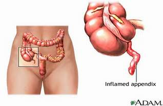
Appendix inflamed and had edema as a result of congestion, possibly by fecalith (hard mass of feces), tumor or a foreign object. Inflammatory process, increased intraluminal pressure that will impede lymph flow resulting in edema, diapedesis bacteria and ulceration of the mucosa cause upper abdominal pain or severe diffuse progressively, within a few hours, localized to the right lower quadrant of the abdomen. Finally, the inflamed appendix contains pus.

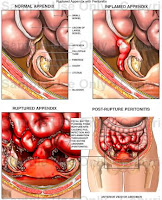
When mucus secretion continues, the pressure will continue to rise causing widespread inflammation and the resulting local peritoneum, causing pain under the right side is called acute suppurative appendicitis. If then the flow will be disrupted arterial wall infarction followed by a gangrenous appendix called gangrenous appendicitis. If the walls are already fragile perforated appendicitis rupture will occur. If all of the above process is slow, omentum and adjacent bowel will move toward an appendix to arise a local mass dsebut appendicular infiltrates. Inflammation of the appendix may be an abscess or disappear.

In children, shorter omentum and appendix are longer, thinner wall of the appendix. The situation is coupled with immune system becomes less ease of perforation. In older people perforation easily happen because there is an interruption of blood vessels (Mansjoer, 2000).

Clinical Manifestations of Appendicitis
  • Lower quadrant pain
  • Mild fever
  • Nausea and vomiting
  • Loss of appetite
  • Local tenderness at the point mc Burney
  • Tenderness off (or intesifikasi result of pain when pressure is released)
  • Signs rovsing can arise by doing palpoasi lower left quadrant which paradoksimal cause pain felt in the lower right quadrant
  • Abdominal distension due to paralytic ileus
  • The patient's condition deteriorates

Nursing Diagnosis Decreased Cardiac Output for Hyperthyroidism

Decreased Cardiac Output related to uncontrolled hyperthyroidism, hypermetabolism, increased cardiac workload.

Patients objective / evaluation criteria;
  • Maintain adequate cardiac output according to the needs of the body

characterized by:
  • Stable vital signs,
  • normal peripheral pulses,
  • normal capillary refill,
  • good mental status,
  • no dysrhythmias

Nursing Intervention:

Independent
  • Monitor vital signs. Note the magnitude of the pressure pulse.
  • Check / meticulous possibility complained of chest pain patients.
  • Assess pulse / heart rate while the patient sleeps.
  • Auscultation of heart sounds, note the extra heart sounds, a gallop rhythm and a systolic murmur.
  • ECG monitor, record or note rate or in cardiac rhythm and the presence of dysrhythmias
  • Observation of signs and symptoms of severe thirst, dry mucous membranes, weak pulse, slow capillary refill, decreased urine output, and hypotension
  • Note adnya history of asthma / bronkokontriksi, pregnancy, sinus bradycardia / heart block progress to heart failure
Collaboration
  • Give fluids through IV as indicated
  • Give medications as indicated:
  • Monitor the results of lab tests: serum potassium, serum calcium, sputum culture
  • Perform regular ECG monitoring
  • Give oxygen as indicated
  • Prepare for surgery

Meningitis Definition, Etiology and Clinical Manifestations

Definition of Meningitis

Meningitis is inflammation of the meninges (the membranes that surround the brain and spinal cord) and is caused by a virus, bacteria or fungus organs (Smeltzer, 2001).

Meningitis is an infection of the fluid of the brain with inflammatory piamater, arachnoid and in a milder degree of the brain and spinal cord tissues were superficial. (Neurology capita selekta, 1996)

Meningitis is an inflammation of the arachnoid and pia mater (lepto meningens) of the brain and spinal cord. Bacteria and viruses are the most common cause of meningitis, although fungi can also cause. Bacterial meningitis is more common. Early detection and treatment will give more better results according to Revelation Widagdo et al (2008:105)

Etiology of Meningitis

Meningitis caused by a virus is generally harmless, will recover without specific treatment and care. But bacterial meningitis can lead to serious conditions, such as brain damage, hearing loss, lack of ability to learn, can even cause death. While meningitis is caused by a fungus is very rare, this type generally affects people with damaged immune (immune system) as in patients with AIDS.

Bacteria that can cause meningitis attack include:
1. Streptococcus pneumoniae (pneumococcus).
These bacteria are the most common cause of meningitis in infants or children. This type of bacteria can also cause pneumonia, ear and nasal cavity (sinus).

2. Neisseria meningitidis (meningococcus).
This bacterium is the second most after Streptococcus pneumoniae meningitis caused by an infection of the upper respiratory tract and then the bacteria enter the bloodstream.

3. Haemophilus influenzae (Haemophilus).
Haemophilus influenzae type b (Hib) is a type of bacteria that can also cause meningitis. This type of virus as the cause upper respiratory infections, middle ear and sinuses. Vaccine (Hib vaccine) has shown a decrease in the number of cases of meningitis caused by these bacteria.

4. Listeria monocytogenes (listeria).
This is one type of bacteria that can cause meningitis. These bacteria can be found in many places, in the dust and in contaminated food. Food is usually a type of cheese, hot dogs and bacon sandwich which is derived from the bacterium local animal (pet).

5. Other bacteria that can also cause meningitis are Staphylococcus aureus and Mycobacterium tuberculosis.

Clinical Manifestations of Meningitis
  • Early in the disease, fatigue, changes in power to remember, change in behavior
  • In accordance with the rapid course of the disease the patient becomes stuporous
  • Headache
  • Sore muscle pain
  • Pupillary reaction to light. Photofobia when light is directed at the patient's eye.
  • Dysfunction of the nerves III, IV, VI
  • Motor movement at the beginning of the disease is usually normal and common in the advanced stages of hemiparesis, hemiplagia, and decreased muscle tone
  • Reflex positive Brudzinski and Kernig reflex
  • Nausea
  • Vomiting
  • Tachycardia
  • Convulsions
  • Patients feel fear and anxiety

Risk for Injury - Nursing Care Plan Meningitis


Nursing Diagnosis for Meningitis: Risk for Injury related to general weakness.

The expected outcomes / evaluation criteria pediatric patients: No seizures or comorbidities or other injury.

intervention
a. Monitor the spasms / twitching of the hands, feet and mouth or other facial muscles.
Rational: reflecting on the CNS in general irritation that require immediate evaluation and possible intervention to prevent complications.

b. Provide security for patients by providing assistance on the bed and keep the barrier remained in place and attach the plastic artificial airway or soft rolls and a suction bulb.
Rationale: protect patients when seizures. Note; enter the airway assistance / soft rolls if only jaw relaxation, not forced to enter as his teeth shut and soft tissue will be damaged.

c. Maintain bed rest during the acute phase. Move. Moving with the help of corresponding improvement in the situation.
Rational: reducing the risk of falls / trauma case vertigo, syncope or ataxia.

d. Give medication as indicated as phenytoin (Dilantin), diazepam, phenobarbital.
Rational: an indication for the treatment and prevention of seizures. Records: Phenobarbital may cause respiratory and sedative defresi and mask the signs / symptoms of increased ICP.

Retropharyngeal Abscess Prevention and Treatment

Retropharyngeal abscess Prevention and Treatment

Definition of retropharyngeal abscess

Retrofaringeal abscess is a pus accumulation in the back of the throat tissues.

Cause of retropharyngeal abscess
  • Abscesses are usually caused by streptococcal infection from the tonsils, throat, sinuses, adenoids, nasal or middle ear.
  • Sometimes an injury to the back of the throat caused by thorn fish can also cause abscesses retrofaringeal.
  • Although rare, retrofaringel abscess can also be caused by tuberculosis.
  • Retrofaringeal abscess usually affects children aged less than 5 years.
  • Networking on the back of the throat allows the formation of children's cavity containing pus (which this does not occur in adults).
  • Infection in these areas can occur during or shortly after by a bacterial throat infection.

Symptoms of retropharyngeal abscess

Symptoms include:
  • History of sore throat, nasal infection or tooth abscess
  • High fever
  • Severe sore throat
  • Swollen neck lymph nodes
  • Difficulty swallowing
  • Salivating
  • Respiratory Disorders
  • Intercostal retraction (withdrawal of muscles between the ribs when people try so hard to breathe)
  • Stridor (harsh breathing sound).

Complications of retropharyngeal abscess
  • Bleeding around the abscess
  • Rupture of the abscess into the airways (which can cause airway obstruction)
  • Pneumonia
  • The spread of infection to the chest.
Diagnosis of retropharyngeal abscess
  • Diagnosis is based on symptoms and physical examination.
  • X-rays or a CT scan of the neck showed a cavity containing pus between the throat and cervical spine.
  • Blood tests showed an increase in the number of white blood cells.
  • Breeding throat mucus can indicate the presence of the causative organism.

Treatment of retropharyngeal abscess
  • To overcome infection drainage surgery (to remove the pus) and high doses of antibiotics given intravenously.

Prevention of retropharyngeal abscess
  • Diagnosis and treatment in pharyngitis and nasopharyngeal infection usually can prevent abscess retrofaringeal.

Physical Examination, Laboratory, and Radiology for Acute Appendicitis

Physical Examination, Laboratory, and Radiology for Acute Appendicitis

There are several tests that can be performed by a medical team to determine and diagnose the presence of Appendicitis by patients. Among them is the physical examination, laboratory tests and radiology examinations:

1. Physical Examination of Acute Appendicitis

Physical Examination of Acute Appendicitis

In acute appendicitis, the observation would seem the swelling of the abdominal cavity which seemed to tighten the abdominal wall (distended). On palpation of the right lower abdominal area, often when pressed will feel pain when pressure is released and also will feel pain (Blumberg sign), which is the key to the diagnosis of acute appendicitis.

By the action of the right leg bent and thighs strong / leg high in the lift, the abdominal pain is getting worse. Suspicion of an inflammation of the appendix is ​​enhanced when an anal or vaginal and cause pain as well. Rectal temperature (rectal) temperatures are higher than the armpits, over longer support the existence of appendicitis.

2. Laboratory Examination of Acute Appendicitis

In laboratory tests of blood, which can be found is an increase of white blood cells (leukocytes) to about 10,000 - 18.000/mm3. If an increase is more than that, then it is likely the appendix had been perforated (ruptured).

3. Radiological Examination of Acute Appendicitis

Radiological Examination of Acute Appendicitis

Plain abdomen may show a fecalith. However, this examination is rarely helpful in diagnosing appendicitis. Ultrasonography (USG) is quite helpful in the diagnosis of appendicitis enforcement (71-97%), especially for pregnant women and children. The highest level of accuracy is by CT scan (93-98%). With a CT scan can be seen clearly picture the appendix.

Risk for Infection - Nursing Care Plan for Appendicitis

Risk for Infection - Nursing Care Plan for Appendicitis
Risk for Infection Appendicitis

Nursing Diagnosis Interventions for Appendicitis: Risk for Infection

Risk factors include:
  • Inadequate primary defense, perforation / rupture of the appendix; peritonitis; abscess formation.
  • Invasive procedures, surgical incisions.
Intervention:

Independent:
  • Monitor vital signs noticed fever, chills, sweating, mental changes, increased abdominal pain.
  • Do a good hand washing and aseptic wound care. Provide complete care.
  • See incision and bandage. Write down the characteristics and wound drainage / drain (if included), the erythema.
  • Provide appropriate information, be honest with the patient / parent close.
Collaboration
  • Take for example the drainage when indicated.
  • Give antibiotics, are as indicated.

Clinical Manifestations of Appendicitis

Clinical Manifestations of Appendicitis

  • Right lower quadrant pain felt and is usually accompanied by mild fever, nausea, vomiting, and loss of appetite.
  • Local tenderness at the point Mc.burney, when done pressure.
  • Tenderness may be encountered off.
  • The degree of muscle spasm and tenderness if there is constipation or diarrhea are not depending on the severity of the infection and the location of the appendix.
  • If the appendix behind the caecum circular, pain and tenderness can feel the lumbar region: when the tip is in the pelvis, these signs can be detected only by examination of the rectal examination.
  • Pain on defecation show ends appendix is near the rectum; pain during urination indicates that the tip of the appendix is close to the bladder or ureter.
  • There is stiffness in the bottom of the right testis muscles can occur.
  • Signs Rovsing can arise with left lower quadrant palpation paradoxical that causes pain felt in the lower right quadrant. If paralytic ileus, and the patient's condition deteriorated.
  • In elderly patients, the signs and symptoms of appendicitis can vary greatly. The signs can be very dubious, suggesting destruction of the bowel or other disease processes. Patients may have no symptoms until he suffered a ruptured appendix. The incidence of perforation of the appendix is higher in the elderly, because many of these patients do not seek medical care as soon as patients are younger.

Clinical Manifestations of Acute Gastritis and Chronic Gastritis

Clinical Manifestations of Acute Gastritis and Chronic Gastritis
Acute Gastritis and Chronic Gastritis

Clinical Manifestations of Acute Gastritis and Chronic Gastritis

Acute Gastritis

Acute Gastritis is very varied, ranging from very mild, asymptomatic, up to very heavy, which can lead to death. In the case of very heavy, very prominent symptoms are:
  1. Hematemetis and melena that can last a very great shock to occur due to blood loss.
  2. In most cases, symptoms are very mild and even asymptomatic. Complaints such as pain that arises in the gut, usually mild and can not be appointed to the appropriate location.
  3. Sometimes accompanied by nausea and vomiting.
  4. GI bleeding is often the only symptom.
  5. In the case of very light bleeding, manifests as occult blood in the stool and will be found in physical signs of deficiency anemia with no apparent etiology.
  6. On physical examination found no abnormalities except usually those with severe bleeding, causing signs and symptoms of hemodynamic disorders as real as hypotension, pallor, cold sweat, tachycardia until the disturbance of consciousness.
Chronic Gastritis
  1. Varied and unclear.
  2. Feeling of fullness, anorexia.
  3. Epigastric distress that is not real.
  4. Early satiety.

Pathophysiology of Chronic Gastritis

Pathophysiology of Chronic Gastritis
Chronic gastritis can be classified as type A or type B.

Type A (often referred to as autoimmune gastritis) resulting from changes in parietal cells, leading to atrophy and cell infiltration. It is associated with autoimmune diseases, such as pernicious anemia and occurs in the fundus or corpus of the stomach.

Type B (sometimes referred to as Helicobacter pylori, also known as H. pylori) is associated with the bacteria H. pylori, dietary factors such as heat or spicy drink, use drugs and alcohol, smoking or reflux of intestinal contents into the stomach. H. Pylori includes bacteria that are not acid resistant, but this kind of bacteria are able to secure himself in the mucosal lining of the stomach. The presence of this bacterium in gastric mucosa of the stomach lining causing weakening and brittle so that stomach acid can penetrate the layer. Thus both gastric acid and the bacteria causing wound or ulcer. The immune system will respond to bacterial infection H. Pylori by sending beads of leukocytes, killer T cells, and other infection-fighting. However, it is not able to fight infection H. The reason pylori can not penetrate the stomach lining. But also can not be removed so that the immune response continues to increase and grow. Polymorph die and release of superoxide radicals damaging compounds in the cells lining the stomach. Extra nutrients are sent to strengthen the leukocyte cells, but nutrition is also a source of nutrients for H. Pylori. Finally, the state of the damaged epithelium of the stomach, forming a superficial ulceration and can cause hemorrhage (bleeding). Within a few days gastritis and even peptic ulcers are formed.

Acute Pain related to Constipation

Acute Pain related to Constipation
Acute pain related to Constipation

Nursing Diagnosis for Constipation : Acute Pain related to the accumulation of hard stool in the abdomen

Goal:
  • Show the pain has diminished

Expected outcomes are:
  • Shows relaxation techniques, individually effective to achieve comfort.
  • Maintain the level of pain on a small scale
  • Reported physical and psychological health.
  • Recognize the causes and the use of measures to prevent pain.
  • Using action to reduce the pain with analgesics and non-analgesics as appropriate.
Nursing Intervention for Acute Pain - NCP Constipation:
Independent:
  • Help the patient to focus more on activities rather than pain, to make of switching via television or radio.
  • Note that the elderly have increased sensitivity to the analgesic effects of opiates.
  • Consider the possibility of drug-drug interactions and drug disease in the elderly.
Rational:
  • Clients can distract from pain.
  • Be careful in giving anlgesik opiates.
  • Be careful in the provision of drugs in the elderly.
Observation:
  • Ask the patient to assess pain or lack of comfort on a scale of 0-10.
  • Use the pain flow sheet.
  • Perform a comprehensive pain assessment.
Rational:
  • Knowing the client's level of pain experienced.
  • Knowing the characteristics of the pain.
  • Knowing the specific pain.

Health education
  • Instruct patient to inform the nurse, if the reduction of pain, less is reached.
  • Provide information about the pain.
Rational:
  • Nurses can perform the right actions, overcoming the client's pain.
  • So that patients do not feel anxious.

Imbalanced Nutrition Less Than Body Requirements related to Loss of Appetite

Imbalanced Nutrition Less Than Body Requirements related to Loss of Appetite
Nursing Care Plan

Imbalanced Nutrition Less Than Body Requirements related to loss of appetite

Goal:
  • Showed good nutritional status.

Expected outcomes are:
  • Tolerance to dietary needs.
  • Retain lean body mass and body weight within normal limits.
  • Laboratory values ​​within normal limits.
  • Reported adequacy of energy levels.
Imbalanced Nutrition Less Than Body Requirements related to loss of appetite - Intervention:
Independent:
  • Create a meal plan with the patient, to put in a feeding schedule.
  • Support family members to bring the patient's favorite food from home.
  • Large portions of food offered during the day when a high appetite.
  • Make sure the diet meets the needs of the body as indicated.
  • Make sure the patient's diet is liked or disliked.
  • Monitor input and expenditure and body weight periodically.
  • Assess the patient's skin turgor.
Rational:
  • Keeping the diet of patients so that patients eat regularly.
  • Patients feel comfortable with food brought from home and can improve the appetite of the patient.
  • By administering a large portion can maintain adequacy of nutrition intake.
  • High carbohydrate, protein, and calories needed or required during treatment.
  • To support the increasing appetite of the patient.
  • Knowing the balance of intake and expenditure of food intake.
  • As the data supporting a change of less nutritional needs.
Observation
  • Monitor laboratory values​​, such as hemoglobin, albumin, and blood glucose levels.
  • Teaches a method for meal planning.
Rational:
  • In order to determine the level of Hb content deficiencies, albumin, and glucose in the blood.
  • Clients used to eat in a planned and orderly.

Health Education
  • Teach patients and families about nutritious food and not expensive.
Rational:
  • Maintain the adequacy of intake of nutrients needed.

Constipation related to the irregular pattern of defecation

Constipation related to the irregular pattern of defecation
Nursing Care Plan for Constipation

Constipation related to the irregular pattern of defecation

Objectives:
Patients can defecate regularly (every day)

Expected outcomes are:
  • Defecation can be done once a day.
  • The consistency of soft stool.
  • Elimination of excess stool without straining.
Intervention:
Independent:
  • Determine the pattern of defecation for clients and train clients to do.
  • Set the time for clients such as defecation after meals.
  • Give the range of fibrous nutrients according to the indication.
  • Give fluids if not contraindicated 2-3 liters per day.
Collaboration:
  • Giving a laxative or enema as indicated.
Rational:
  • To restore order to the client defecation pattern.
  • To facilitate the defecation reflex.
  • High fiber nutrients to carry fecal elimination.
  • To soften the stool elimination.
  • To soften the stool.

2 Nursing Diagnosis and Interventions for Abdominal Typhoid

2 Nursing Diagnosis and Interventions for Abdominal Typhoid
Nursing Diagnosis and Interventions for Abdominal Typhoid

Nursing Care Plan for Abdominal Typhoid : 2 Nursing Diagnosis and Interventions

1. Imbalanced Nutrition Less Than Body Requirements related to no appetite, nausea and bloating.

Purpose: Improve the nutritional and fluid needs.

Nursing Interventions:
  • Assess nutritional status of children.
  • Allow children to foods that can be tolerated child, plan to improve the nutritional quality at the child's appetite increases.
  • Give the food is accompanied by a nutritional supplement to improve the quality of nutritional intake.
  • Encourage parents to provide food to the technique of small but frequent portions.
  • Weigh weight every day at the same time and with the same scale.
  • Maintain cleanliness of the child's mouth.
  • Explain the importance of adequate nutritional intake for the cure of disease.
  • Collaboration for parenteral feeding through. If feeding via oral did not meet the nutritional needs of children.

2. Risk for Fluid Volume Deficit related to the lack of fluid intake and increased body temperature.

Purpose: To prevent the lack of fluid volume.

Nursing Interventions:
  • Observation of vital signs (body temperature) at least every four hours.
  • Monitor the increasing signs of dehydration: inelastic turgor, sunken fontanel, decreased urine output, dry mucous membranes, cracked lips.
  • Observation and record intake and output and maintain an adequate intake and output.
  • Monitor and record the weight at the same time and with the same scale.
  • Monitor the provision of intravenous fluids through an IV every hour.
  • Reduce the loss of fluid that is not visible (insensible water loss / IWL) to give a cold compress or a tepid sponge.
  • Give antibiotics according to the program.

Nursing Management of Varicella

Nursing Management of Varicella

Nursing Management of Varicella

Patients should be isolated from others, as well as for their daily needs. such as:
  • Isolation to prevent transmission.
  • Nutritious diet (high in calories and protein).
  • If a high fever, compress with warm water.
  • Strive to avoid infection of the skin such as the provision of antiseptic in the water.
  • Strive to vesicles did not rupture
    • Do not scratch the vesicles.
    • Long nails should not be allowed.
    • If want to dry off, just a towel on the skin and do not rub.

The provision of drugs to reduce symptoms such as itching, fever, weight is required in order to reduce the level of disease. Antiviral drug, is recommended within 48 hours of patients complaining of symptoms of varicella.

Giving varicella zooster immuno-globulin (VZIG) is given less than 96 hours after exposure, namely on:
  • Women with pregnancy.
  • Children with impaired immune systems.
  • Newborns whose mothers contracted varicella within 5 days before delivery or 48 hours after birth.
  • Premature infants 28 weeks of age or younger with a parent without a history of previous varicella.

Nursing Diagnosis for Pharyngitis

Nursing Diagnosis for Pharyngitis

Pharyngitis

Pharyngitis is the commonest cause of simple "sore throat". It may be due to infective or other causes. Viral infections include coryza, influenza, adenovirus, herpes simplex, measles, rubella, chickenpox, Coxsackie and echoviruses. Bacterial pathogens such as streptococci, Staphylococci, Penumococci, H. Influenzae, C. diphtheriae, fusobacterium, gonococci and T. Pallidum and fungi such as Candida albicans may lead to pharyngitis. In agranulocytosis and acute leukemia, necrotic ulceration of the tonsils and pharynx may be the presenting symptom. Excessive smoking, noxious fumes, corrosive and unaccustomed spicy foods are common irritants. Pharyngitis may also be caused by allergy to inhaled or ingested allergens.


Nursing Diagnosis for Pharyngitis

1. Acute pain related to inflammation of the throat.

2. Ineffective airway clearance related to thick secretions characterized by difficulty in breathing.

3. Imbalance nutrition less than body requirements related to difficulty swallowing.

4. Knowledge Deficit related to not familiar with the sources of information.

Pathophysiology of Pharyngitis

Pathophysiology of Pharyngitis

Pathophysiology of Pharyngitis

Transmission is by droplet. Germs infiltrate the epithelial layer, then the epithelium is eroded, then the superficial lymphoid tissue inflammatory reaction occurs with the damming of polymorphonuclear leukocyte infiltration. In the early stages there is hyperemia and edema and increased secretions. Serous exudate at first, but tends to become thickened and dry and can be attached to the pharyngeal wall. By hyperemia, blood vessel walls of the pharynx to the width. Blockage forms a yellow, white, or gray present in follicles or lymphoid tissue. It appears that the lymphoid follicles and blotches on the posterior pharyngeal wall or located more laterally to become inflamed and swollen causing inflammation of the throat or pharyngitis.

NCP Stroke: Ineffective Cerebral Tissue Perfusion related to interruption of blood flow

NCP Stroke: Ineffective Cerebral Tissue Perfusion related to interruption of blood flow

NCP Stroke: Ineffective Cerebral Tissue Perfusion related to interruption of blood flow

Ineffective Cerebral Tissue Perfusion related to interruption of blood flow: occlusive disease, hemorrhage, cerebral vascular spasm, cerebral edema.

Expected outcomes are:
  • Maintained and increased the level of consciousness, cognition and function of sensory / motor.
  • Reveals stabilization of vital signs and no increase in intracranial pressure.
  • The role of the patient reveals no deterioration / recurrence.

Nursing Intervention:

Independent
  • Determine the factors related to individual situations / causes of coma / decreased cerebral perfusion and a potential increase in intracranial pressure.
  • Monitor and record neurological status on a regular basis.
  • Monitor vital signs.
  • Pupil evaluation (size, shape similarity and reaction to light).
  • Help to change the view, misalnay blurred vision, visual field changes / perceptual field of view.
  • Increasing auxiliary functions, including speech if the patient has impaired function.
  • And gradual elevation of the head in neutral position.
  • Maintain bed rest, provide a quiet environment, set visits as indicated.
  • Provide supplemental oxygen as indicated.
  • Give medications as indicated.

Medical Management of Scoliosis

Medical Management of Scoliosis

Medical Management of Scoliosis

Treatment depends on the cause, degree and location of the curve and the stage of bone growth.

If the curvature is less than 20%, usually require no treatment, but patients should perform regular checks every 6 months.

In children who are still growing, the curvature usually increased up to 25-30%, because it is usually advisable to use braces to help slow the progression of the curvature of the spine.

Braces of the "Milwaukee and Boston" is effective in controlling the progression of scoliosis, but must be installed for 23 hours / day until the child stops growing.
Brace ineffective in congenital and neuromuscular scoliosis.

If the curvature reached 40% or more, usually with surgery. In the surgery done to improve the curvature and fusion of the bones. Bone is maintained in place with the help of 1-2 metal tool attached to the bones recovered (less than 20 years). After the surgery may need to be fitted brace to stabilize the spine.

Sometimes given elektrospinal stimulation, in which the spinal muscles were stimulated with a low electrical current to straighten the spine.

Pathophysiology of Scoliosis

Pathophysiology of Scoliosis

Pathophysiology of Scoliosis

Deformity of the spine called scoliosis, originated from the nerves of weak or even paralyzed attractive segments of the spine. This serves to keep the pull of the vertebrae are in the normal lines of its shape like a ruler or straight. But for some reason, such as the habit of sitting the oblique, make the most of the work to be weak nerves. If this continues over and over becomes a habit, then the nerve will even die. This results in an imbalance of pull on the vertebrae. Therefore, suffering from scoliosis of the spine is curved like the letter or the letter S or C.

Nursing Assessment for Scoliosis

Nursing Assessment for Scoliosis

Nursing Care Plan for Scoliosis : Nursing Assessment for Scoliosis

The physical examination includes:

a. Assessing the body's skeletal
The presence of deformity and alignment. Abnormal bone growth due to bone tumors. Shortening of the extremities, amputation and body parts that are not in anatomic alignment. Abnormal angulation of the long bones or motion at a point other than the joints usually indicate a fracture.

b. Assessing the spine
Scoliosis (lateral curvature of the spine deviation)

c. Assessing the joint system
Extensive movement are evaluated either actively or passively, deformity, stability, and bruising, stiffness of joints.

d. Assessing the muscle system
The ability to change position, muscle strength and coordination, and the size of each limb to mementau otot.Lingkar edema or atropfi, muscle pain.

e. Examine how to walk
The existence of irregular movements are not considered normal. If one limb shorter than the others. A variety of neurological conditions associated with abnormal gait (eg walking spastic hemiparesis - stroke, how to go step by step - lower motor neuron disease, how to walk vibrate - Parkinson's disease).

f. Examine the skin and peripheral circulation
Palpation of the skin may indicate a yanglebih temperature hotter or colder than others and adanyaedema. Peripheral circulation was evaluated by assessing peripheral pulses, color, temperature and capillary refill time.

Analysis of data
Subjektif Data :
  • Back pain patients say
  • Patients said fatigue in the spine after sitting or standing for long
  • Patients say trouble breathing

Objective Data :
  • That looks are not the same shoulder height
  • Visible protrusion of the scapula is not the same
  • Looks are not the same hip

9 Tips to overcome nausea and vomiting during pregnancy

9 Tips to overcome nausea and vomiting during pregnancy

Tips to overcome nausea and vomiting during pregnancy

Some tips to help you cope with "morning sickness" or nausea-vomiting during early pregnancy:

A. Eat small amounts, but often, do not eat in large quantities or portions, will only make you grow sick. Try to eat when you should eat small meals but frequently.

2. Eating foods high in carbohydrates and protein that can help overcome your nausea. Many fruit and vegetables and foods high in carbohydrates such as bread, potatoes, biscuit, etc..

3. In the morning when you wake up, do not jump to hasty wake up, try to sit first, and only slowly stood up. If you feel very sick when you wake up in the morning prepare snack or biscuit near your bed, and you can eat it before you try to stand.

4. Avoid fatty foods, oily and spicy foods that will aggravate your nausea.

5. Drink enough to avoid dehydration from vomiting. Drink water, or juice. Avoid drinks that contain caffeine and carbonates.

6. Prenatal vitamins sometimes exacerbate nausea, but you still need folate for pregnancy is. If nausea and vomiting is very severe, consult your doctor so that it can be given the best advice for vitamins that you will consume. And your doctor will probably provide a cure for nausea when necessary.

7. Vitamin B6 is effective for reducing nausea in pregnant women. Should first consult with your doctor to use.

8. Traditional Treatment: Usually, people use ginger in reducing nausea in a variety of traditional medicine. Research suggests that ginger can be used as traditional medicine to relieve nausea and safe for mother and baby. At some pregnant women who consume fresh ginger or ginger candy to help cope with her nausea.

9. Rest and relax will help you cope with nausea vomiting. Because if you stress will only aggravate your nausea. Uptake time for you! just try to rest and relax, listen to music, reading books or magazines of your favorite baby etc.. Deal with your pregnancy with happiness, because it is grace. :-)

Remember! Call your doctor if nausea and vomiting become so severe, so that you can not eat or drink anything that can lead to lack of fluids / dehydration.

Believe Morning sickness or nausea and vomiting in early pregnancy it will pass without you realizing it and this will be one exciting experience during your pregnancy, just think about the little one will be coming soon bring happiness million.

Nursing Diagnosis for Impetigo

Nursing Diagnosis for Impetigo
Nursing Care Plan for Impetigo Nursing Diagnosis for Impetigo

Nursing Diagnosis for Impetigo

Impetigo is a highly contagious bacterial skin infection most common among pre-school children. People who play close contact sports such as rugby, American football and wrestling are also susceptible, regardless of age. Impetigo is not as common in adults. The name derives from the Latin impetere ("assail"). It is also known as school sores.

It is primarily caused by Staphylococcus aureus, and sometimes by Streptococcus pyogenes. According to the American Academy of Family Physicians, both bullous and nonbullous are primarily caused by Staphylococcus aureus, with Streptococcus also commonly being involved in the nonbullous form.


Nursing Diagnosis for Impetigo

1. Impaired Skin Integrity
2. Risk for Infection
3. Disturbed Body Image
4. Anxiety
5. Knowledge Deficit

Nursing Diagnosis for Congenital Dislocation Of The Hip

Nursing Diagnosis for Congenital Dislocation Of The Hip
Nursing Care Plan for Congenital Dislocation Of The Hip - Nursing Diagnosis for Congenital Dislocation Of The Hip

Congenital dislocation or subluxation of the hip (congenital acetabular dysplasia) is a complete or partial displacement of the femoral head out of the acetabulum. The physical signs are essential for the diagnosis of age related. In newborns the tests for instability are the most sensitive. After the neonatal period, and until the age of walking, tightness of the adductor muscles is the most reliable sign. Early diagnosis is vital for Successful treatment of this condition partially genetically determined. Various therapeutic measures, ranging from abduction splinting to open reduction and osteotomy, may be required. Following the diagnosis in the first month of life, the average treatment time in one recent series was only 2.3 months from initiation of therapy to Attainment of a normal hip. When the diagnosis was not made until 3 to 6 months of age, ten months of treatment was required to Achieve the same outcome. When the diagnosis is not made, or the treatment is not Begun until after the age of 6, a normal hip will probably not develop in any patient. (ncbi.nlm.nih.gov)

Nursing Diagnosis for Congenital Dislocation Of The Hip
  1. Acute Pain related to dislocation
  2. Impaired Physical Mobility related to pain during mobilization
  3. Disturbed Body Image related to changes in body shape

4 Nursing Diagnosis for Scoliosis

4 Nursing Diagnosis for Scoliosis
Nursing Care Plan for Scoliosis Nursing Diagnosis for Scoliosis

Nursing Diagnosis for Scoliosis

Scoliosis affects 2% of women and 0.5% of men in the general population. There are many Causes of scoliosis, congenital spine deformities Including, genetic conditions, neuromuscular problems and limb length inequality. Other Causes for scoliosis include cerebral palsy, spina bifida, muscular dystrophy, spinal muscular atrophy and tumors. Over 80% of scoliosis cases, however, are idiopathic, the which means That there is no known cause. Most idiopathic scoliosis cases are found in otherwise healthy people.

Symptoms

Scoliosis can be mild, moderate or severe. The symptoms and signs of scoliosis can include:
  • One shoulder tilted down towards a raised hip, as if the child is Leaning sideways
  • Prominent ribs
  • A protruding shoulder blade
  • Tilted waist
  • The curve is more pronounced when the child bends forward.

4 Nursing Diagnosis for Scoliosis
  1. Ineffective Breathing Pattern
  2. Acute Pain
  3. Impaired Physical Mobility
  4. Disturbed Body Image
Source : http://nandanursingdiagnoses.blogspot.com/2012/07/nursing-diagnosis-scoliosis.html

    Impaired Physical Mobility Nursing Care Plan Scoliosis

    Impaired Physical Mobility Nursing Care Plan Scoliosis
    Impaired Physical Mobility Nursing Care Plan Scoliosis

    Impaired Physical Mobility Nanda Definition: a nursing diagnosis approved by the North American Nursing Diagnosis Association, defined as the state in the which an individual has a Limitation in independent, purposeful physical movement of the body or of one or more extremities.


    Alteration in mobility may be a temporary or more permanent problem. Most disease and rehabilitative states involve some degree of immobility (eg, as seen in strokes, leg fracture, trauma, morbid obesity, and multiple sclerosis). With the longer life expectancy for most Americans, the incidence of disease and disability Continues to grow. And with shorter hospital stays, Patients are being transferred to rehabilitation facilities or sent home for physical therapy in the home environment.

    Mobility is also related to body changes from aging. Loss of muscle mass, reduction in muscle strength and function, stiffer and less mobile joints, affecting balance and gait changes can significantly compromise the mobility of elderly Patients. Mobility is paramount if elderly Patients are to maintain any independent living. Restricted movement affects the performance of most activities of daily living (ADLs). Elderly Patients are also at Increased risk for the complications of immobility. Nursing goals are to maintain functional ability, Prevent additional impairment of physical activity, and Ensure a safe environment.

    Nursing Diagnosis for Scoliosis: Impaired Physical Mobility related to postural imbalance.

    Purpose : Increase physical mobility

    Plan of action
    1. Assess the level of physical mobility.
    Rational: Influencing choice / control the effectiveness of interventions.

    2. Increase activity if pain is reduced.
    Rationale: Provides the opportunity to release energy.

    3. Teaching aids and active joint range of motion exercises.
    Rationale: Increases muscle strength and circulation.

    4. Involve the family in performing self-care.
    Rational: The family that cooperate to relieve the officer, and provide comfort to patients.

    Nursing Diagnosis Risk for Infection - Tuberculosis NCP

    Nursing Diagnosis Risk for Infection - Tuberculosis NCP
    Nursing Diagnosis Risk for Infection

    Nursing Care Plan for Tuberculosis

    Nursing Diagnosis : Risk for Infection related to lack of knowledge in order to avoid exposure of pathogens.

    Expected outcomes are:
    • Lowers the risk of spreading infection

    Nursing Interventions Risk for Infection - Tuberculosis NCP:
    • Review of pathology of the disease.
    • Identification of others at risk.
    • Instruct patient to cough / sneeze and remove the tissue and avoid spitting.
    • Review of infection control measures.
    • Keep an eye on the temperature as indicated.
    • Collaboration with the medical team.
    Rationalization:
    • Help patients recognize / accept the need to comply with treatment programs.
    • People who are exposed to drug treatment programs to prevent the spread / infection.
    • Can help reduce the patient's sense of isolation.
    • Febrile reaction indicators of infection.
    • Help identify institutions that could be reached to reduce the spread of infection.

    Knowledge Deficit related to Tuberculosis

    Knowledge Deficit related to Tuberculosis
    Nursing Diagnosis Knowledge Deficit related to Tuberculosis

    Nursing Diagnosis and Interventions - Knowledge Deficit related to Tuberculosis

    Nursing Diagnosis for TB Tuberculosis: Knowledge Deficit: about the conditions, actions, and prevention related to inaccurate and incomplete information.

    Expected outcomes are:
    • Said understanding the disease process / prognosis and treatment needs.

    Nursing Interventions Knowledge deficit related to Tuberculosis:
    • Assess the patient's ability to learn.
    • Identification of symptoms should be reported to the nurse.
    • Provide written instructions and information.
    • Encourage clients not to smoke.
    • Assess how TB is transmitted
    Rationalization:
    • Learning depends on emotional and physical readiness and improved at the individual stages.
    • Can show progress or reactivation of disease or drug effects that require further evaluation.
    • Written information for patients given the lower barriers to large amounts of information.
    • Although smoking does not stimulate the recurrence of TB but increased respiratory dysfunction.

    Ineffective Airway Clearance related to Tuberculosis

    Ineffective Airway Clearance related to Tuberculosis

    Ineffective Airway Clearance related to Tuberculosis

    Ineffective Airway Clearance related to Tuberculosis

    Nursing Diagnosis for TB Tuberculosis: Ineffective airway clearance related to thick secretions or blood secretions.

    Expected outcomes are:
    • Maintain the patient's airway
    • Removing secretions without help


    Nursing Interventions Ineffective Airway Clearance related to Tuberculosis:

    • Assess respiratory function, eg, breath sounds, speed, rhythm, depth and use of accessory muscles.
    • Note the ability to remove mucous / coughing effective: note the character, amount of sputum, presence of hemoptysis.
    • Give the patient or the semi-Fowler position higher. Help the patient to cough and deep breathing exercises.
    • Clean secretions from the mouth and trachea: suction as needed.
    • Collaboration with the medical team in the provision of drugs.
    Rrationalization:
    • Decrease in breath sounds may indicate atelectasis.
    • Expenditure is difficult when the secretions are very thick. Bloody sputum or blood thick bright due to lung damage or injury and may require evaluation bronkal.
    • Position to help maximize lung expansion and reduce respiratory effort.
    • Prevent obstruction / aspiration.

    Prevention of Vaginal Discharge in Women

    Prevention of Vaginal Discharge in Women
    Prevention of vaginal discharge in women

    Prevention of Vaginal Discharge in Women

    Vaginal discharge is a problem that has long been a problem for women. Not many women who know what a vaginal discharge, and sometimes underestimate the problem of vaginal discharge. Though Vaginal discharge can not be taken lightly, as a result of vaginal discharge can be very fatal if treated late.

    Vaginal discharge or Fluor Albus is a condition where excess fluid out of the vagina. Vaginal discharge can be divided into 2 parts whitish normal (physiological) and vaginal discharge abnormal (pathological). Whiteness is not a disease (physiological) can occur on any girl. Discharge is usually clear, colorless, odorless and does not itch. This amount of fluid discharge can be a little or a lot, occurred before and after menstruation, when sexually aroused or when you're stressed. Sometimes we also experience vaginal discharge that teenagers just before puberty, usually the symptoms will go away by itself.

    But if the discharge from the vagina is not clear, yellowish white, gray to greenish, thick, smelling like rotten eggs or rancid, itching and more numerous, it is likely that whiteness is not normal. Some of the causes of abnormal vaginal discharge caused by infection is usually accompanied by itching in the vagina and around the outer vaginal lips. Which often cause vaginal discharge include bacteria, viruses, fungi or parasites also. If not treated the infection can spread and cause inflammation of the urinary tract, causing the pain when the patient is urinating.

    Vaginal discharge can be prevented by:
    • Keeping the genital hygiene, cleaning the vagina with clean running water by wiping from front to back.
    • Minimize the use of antiseptic soap because it can interfere with vaginal pH balance.
    • Time to change pads at least 3 times a day.
    • Choosing the right underwear, not wearing pants that are tight and absorb perspiration.
    • Avoiding risk factors for infection such as sexual promiscuity, as well as regular gynecological examinations.

    Common Causes of Vaginal Discharge

    Common Causes of Vaginal Discharge
    Common Causes of Vaginal Discharge

    Common Causes of Vaginal Discharge

    Vaginal discharge is term given to biological fluids contained within or expelled from the vagina.

    While most discharge is normal and can reflect the various stages of a woman's cycle, some discharge can be a result of an infection, such as a sexually transmitted disease.

    Common Causes of Vaginal Discharge 
    • Often using tissue, while washing the female, after urinating or defecating.
    • Wearing tight underwear from synthetic materials.
    • Often use a dirty toilet.
    • Not change the panty liner.
    • Rinsing the vagina from the wrong direction. Namely from the anus toward the vagina towards the front.
    • Often exchanged briefs / towels with others.
    • Less maintain the cleanliness of the vagina.
    • Exhaustion.
    • Stress.
    • Not immediately replace the pads during menstruation.
    • Wearing any soap to wash the vagina.
    • Not leading a healthy lifestyle (eat irregularly, never exercise, slept less).
    • Living in the humid tropics.
    • Environmental sanitation is dirty.
    • Often with warm water bath and heat. The fungus that causes vaginal discharge is more likely to grow in warm conditions.
    • Frequently change partners in sex.
    • High blood sugar levels.
    • Hormonal imbalance.
    • Frequent scratching vagina.

    Nursing Care Plan for Acute Otitis Media

    Nursing Care Plan for Acute Otitis Media
    Nursing Care Plan for Acute Otitis Media

    Nursing Diagnosis for Acute Otitis Media and Nursing Interventions for Acute Otitis Media

    Nursing Assessment for Acute Otitis Media
    • Assess the presence of pain behaviors: verbal and non-verbal.
    • Assess the increase in temperature (an indication of the infection process).
    • Assess the presence of enlarged lymph nodes in the neck area.
    • Assess nutritional status and adequacy of fluid intake of calories.
    • Assess the possibility of deafness.

    Nursing Diagnosis for Acute Otitis Media
    1. Acute Pain related to inflammation of the middle ear tissue.
    2. Disturbed Sensory Perception: auditory conductive disorder related to the sound of the organ.

    Nursing Interventions for Acute Otitis Media

    1. Acute Pain related to inflammation of the middle ear tissue.

    Purpose: The reduction in pain.

    Intervention:
    • Assess the level of intensity of the client and client's coping mechanisms.
    • Give analgesics as indicated.
    • Distract the patient by using relaxation techniques: distraction, guided imagination, touching, etc..

    2. Disturbed Sensory Perception: auditory conductive disorder related to the sound of the organ.

    Purpose: to improve communication

    Intervention:
    • Reduce noise in the client environment.
    • Looking at the client when speaking.
    • Speaking clearly and firmly on the client without the need to shout.
    • Provide good lighting when the client relies on the lips.
    • Using the signs of non-verbal (eg facial expressions, pointing, or body movement) and other communications.
    • Instruct family or the people closest to the client on how techniques of effective communication so that they can interact with clients.
    • If the client wants, the client can use hearing aids.

    Pathophysiology of Acute Otitis Media

    Pathophysiology of Acute Otitis Media
    Pathophysiology of Acute Otitis Media
    Pathophysiology of Acute Otitis Media

    Acute otitis media is often preceded by respiratory tract infections such as sore throats / colds that spread to the middle ear through the eustachian channel.

    When the bacteria through the eustachian, the bacteria can cause infections of the channel. So that there was swelling around the channel, channel blockage, and the coming of the white blood cells to fight bacteria.

    White blood cells will fight the bacterial cells at the expense of their own, at least to form pus in the middle ear. Tissue swelling around the eustachian cells causes mucus produced when cells multiply mucus and pus, hearing can be impaired because the eardrum and small bones connecting the ear drum with the hearing organ in the inner ear to move freely. Too much fluid, finally able to tear the eardrum because of the pressure.

    Nursing Care Plan for Osteomyelitis

    Nursing Care Plan for Osteomyelitis
    Nursing Care Plan for Osteomyelitis

    Nursing Care Plan for Osteomyelitis : Nursing Diagnosis for Osteomyelitis and Nursing Interventions for Osteomyelitis

    Osteomyelitis is an infection of the bone. It can be caused by a variety of microbial agents (most common in staphylococcus aureus) and situations, including:
    • An open injury to the bone, such as an open fracture with the bone ends piercing the skin.
    • An infection from elsewhere in the body, such as pneumonia or a urinary tract infection that has spread to the bone through the blood (bacteremia, sepsis).
    • A minor trauma, which can lead to a blood clot around the bone and then a secondary infection from seeding of bacteria.
    • Bacteria in the bloodstream bacteremia (poor dentition), which is deposited in a focal (localized) area of the bone. This bacterial site in the bone then grows, resulting in destruction of the bone. However, new bone often forms around the site.
    • A chronic open wound or soft tissue infection can eventually extend down to the bone surface, leading to a secondary bone infection.

    Symptoms of osteomyelitis

    The symptoms of osteomyelitis can include:
    • Pain and/or tenderness in the infected area
    • Swelling and warmth in the infected area
    • Fever
    • Nausea, secondarily from being ill with infection
    • General discomfort, uneasiness, or ill feeling
    • Drainage of pus through the skin

    Additional symptoms that may be associated with this disease include:
    • Excessive sweating
    • Chills
    • Lower back pain (if the spine is involved)
    • Swelling of the ankles, feet, and legs
    • Changes in gait (walking pattern that is a painful, yielding a limp)


    Nursing Diagnosis for Osteomyelitis
    1. Acute pain related to inflammation and swelling
    2. Impaired Physical Mobility related to pain and limitation of the load weight
    3. Risk for Infection

    Targets to be achieved:
    1. Pain is reduced
    2. Improvement of physical mobility within the limits of therapeutic
    3. Infection control

    Nursing interventions for Osteomyelitis

    1. Immobilization of the affected area with a splint to reduce pain and muscle spasms.

    2. Joints above and below the affected area should be made so that still can be moved according to the range yet gently. The wound itself is sometimes very painful and must be handled carefully and slowly.

    3. Elevate the affected area to reduce swelling and discomfort.

    4. Monitor the affected extremity neurovascular status.

    5. Do pain management techniques such as massage, distraction, relaxation, hypnosis to reduce pain perception and collaboration with medical for providing analgesic.

    6. Protect your bones by means of immobilization and avoid stress on the bone because bones become weak due to the infection process.

    Reference : http://my.clevelandclinic.org/disorders/osteomyelitis/hic_osteomyelitis.aspx

    5 Nursing Diagnosis for Tonsillitis

    5 Nursing Diagnosis for Tonsillitis
    Nursing Diagnosis for Tonsillitis

    Nursing Care Plan for Tonsillitis - Nursing Diagnosis for Tonsillitis

    Tonsillitis refers to inflammation of the pharyngeal tonsils.The inflammation may involve other areas of the back of the throat including the adenoids and the lingual tonsils.The tonsils are lymph nodes, or oval-shaped masses of lymph gland tissue, located on both sides of the throat. An infection of the tonsils is called tonsillitis.

    There are several variations of tonsillitis: acute, recurrent, and chronic tonsillitis and peritonsillar abscess. This swelling is usually caused by either a viral or bacterial infection. Tonsillitis is the name given to swollen, red, and tender tonsils.

    Tonsillitis is usually a self-limiting condition, ie it gets better without treatment, and generally there are no complications.Tonsillitis is extremely common in children and young people but it can occur at any age. The characteristics of the disease are pain in the throat and trouble swallowing.

    Tonsillitis usually begins with sudden sore throat and painful swallowing.

    5 Nursing Diagnosis for Tonsillitis

    1. Acute pain related to the presence of inflammation in tosil.

    2 · Imbalanced Nutrition Less Than Body Requirements related to inadequate intake.

    3 · Hyperthermia related to acute infection by microorganisms.

    4 · Disturbed Sleep Pattern related to the pain in the tonsil area.

    5. Anxiety related to a lack of knowledge or information about the illness suffered by the client.

    Nursing Diagnosis and Interventions Anxiety related to CHF

    Nursing Diagnosis and Interventions Anxiety related to CHF

    Nursing Diagnosis and Interventions Anxiety related to CHF

    Nursing Care Plan for CHF

    Nursing Diagnosis: Anxiety related to fear of cardiovascular death, decreased health status, a crisis situation, health changes.

    Goal:
    Overcome anxiety

    Expected outcomes are:
    • The client was calm
    • The client understands about the process of nursing and medicine

    Nursing Intervention:

    · Review the signs of verbal expressions of anxiety.
    Rational: the level of anxiety may develop panic that can stimulate the sympathetic with the release of catecholamines lead to increased cardiac demand for oxygen.

    · Accompany the client during the period of high anxiety, give strength, use it at ease.
    Rational: the sense of empathy is a treatment and may increase the client's coping abilities.

    · Orient the client with routine procedures and activities that are expected.
    Rational: orientation can reduce anxiety.

    · Give the client an opportunity to express his concerns.
    Rational: to eliminate ketegangang to the concerns that are not expressed.

    · Do the approach and communication.
    Rational: to foster mutual trust.

    · Give the opportunity to accompany the person closest to the client.
    Rational: to improve safety on the client.

    · Provide an explanation of the disease, causes and treatment to be performed.
    Rational: to provide assurance about the action steps that will be provided so that clients and families to get clear information.

    Nursing Intervention for Osteoporosis

    Nanda Nursing Diagnosis for Osteoporosis
    1. Chronic pain
    2. Disturbed body image
    3. Self-care deficit
    4. Imbalanced nutrition: Less than body requirements
    5. Impaired physical mobility
    6. Risk for impaired skin integrity
    7. Risk for injury
    Source : http://nandanursingdiagnoses.blogspot.com
      Nursing Outcome for Osteoporosis
      1. Client will experience increased comfort and decreased pain.
      2. Client will express positive feelings about himself.
      3. Client will perform activities of daily living within normal limits.
      4. Client will maintain adequate food intake.
      5. Client will maintain joint mobility and range of motion.
      6. Client will demonstrate integrity intact skin.
      7. Client will show the steps to prevent injury.
      Nursing Intervention for Osteoporosis
      1. Explain all treatments, tests, and procedures. For example, if the patient underwent surgery, explain all procedures and preoperative and postoperative care for patients and their families.
      2. Make sure the client and his family clearly understand the prescribed drug regimen. Tell them how to recognize a significant adverse reactions. Instruct them to immediately report it.
      3. Stressed the need for regular gynecological examinations. Also instructed him to immediately report abnormal vaginal bleeding, to detect the hormone estrogen.
      4. If clients take calcium supplements, encouraging liberal fluid intake to help maintain adequate urine output and thus avoid kidney stones, hypercalcemia, and hypercalciuria.
      5. Tell the client to report the immediate pain, especially after trauma.
      6. Explain kliien and osteoporosis in the family so that they can act to prevent fractures.
      7. Instruct patient to eat foods rich in calcium. Explain that the type II osteoporosis can be prevented with adequate calcium intake and regular exercise. Hormonal and fluoride treatments can also help prevent osteoporosis.
      8. Strengthen the patient's efforts to adapt, and shows how his condition has improved or stabilized. Necessary, refer to an occupational therapist or health care professionals to help with daily activities at home.

      Deficient Fluid Volume related to Peritonitis

      Deficient Fluid Volume related to Peritonitis
      Nursing Diagnosis Deficient Fluid Volume
      Nursing Diagnosis : Deficient Fluid Volume - Nursing Care Plan for Peritonitis


      Deficient Fluid Volume NANDA Definition: Decreased intravascular, interstitial, and/or intracellular fluid. This refers to dehydration, water loss alone without change in sodium

      Peritonitis is an inflammation of the peritoneum, the thin tissue that lines the inner wall of the abdomen and covers most of the abdominal organs. Peritonitis may be localised or generalised, and may result from infection (often due to rupture of a hollow organ as may occur in abdominal trauma or appendicitis) or from a non-infectious process.

      Nursing Interventions Deficient Fluid Volume - Nursing Care Plan for Peritonitis

      Independent:

      1. Monitor vital signs, note the presence of hypotension (including postural changes), tachycardia, tachypnea, fever. Measure CVP if any.
      Rational: To assist in the evaluation of the degree of fluid deficit / effectiveness of fluid replacement therapy and response to treatment.

      2. Maintain adequate intake and output and then connect with the body weight daily.
      Rationale: Demonstrates overall hydration status.

      3. Rehydration / resuscitation fluid
      Rationale: To meet the need of fluid in the body (homeostasis).

      4. Measure specific gravity of urine
      Rationale: Demonstrates changes in hydration status and renal function.

      5. Observation of skin / mucous membranes for dryness, turgor, note peripheral edema / sacral.
      Rational: Hypovolemia, fluid displacement, and lack of nutrition aggravate skin turgor, adding tissue edema.

      6. Eliminate the danger signs / smells from environment. Limit intake of ice cubes.
      Rational: Lowering the gastric stimulation and vomiting response.

      7. Change positions frequently give skin care with often, and keep the bed dry and free of folds.
      Rational: tissue edema and circulatory disturbance tends to damage the skin.

      Collaboration:

      1. Monitor laboratory examinations, eg Hb / hematocrit, electrolytes, protein, albumin, BUN, creatinine.
      Rationale: Provides information about hydration and organ function.

      2. Give the plasma / blood, fluids, electrolytes.
      Rational: Charge / maintain circulating volume and electrolyte balance. Colloid (plasma, blood) to help move the water into the area by increasing intravascular osmotic pressure.

      3. Keep fasting with nasogastric aspiration / intestinal
      Rational: Lowering intestinal hyperactivity, and loss from diarrhea.

      Source : http://careplannursing.blogspot.com/2012/01/deficient-fluid-volume-nursing-care.html
      Copyright © Nursing Diagnosis Intervention. All rights reserved. Template by CB | Published By Kaizen Template | GWFL | KThemes